. 2012 Feb 9;119(6):1468-78.
doi: 10.1182/blood-2011-07-368050. Epub 2011 Nov 16.

P-selectin glycoprotein ligand regulates the interaction of multiple myeloma cells with the bone marrow microenvironment

Affiliations

P-selectin glycoprotein ligand regulates the interaction of multiple myeloma cells with the bone marrow microenvironment

Abdel Kareem Azab et al. Blood. .

Erratum in

Abstract

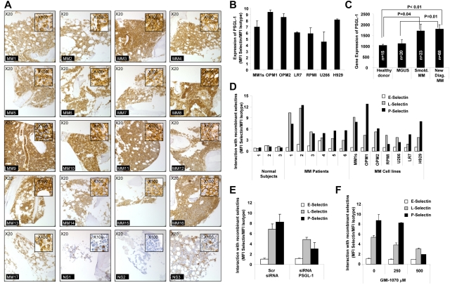
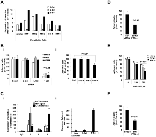
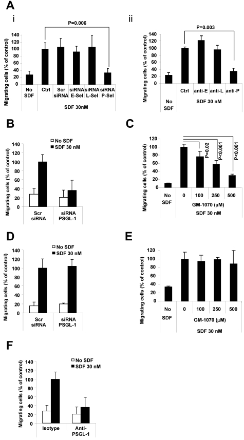
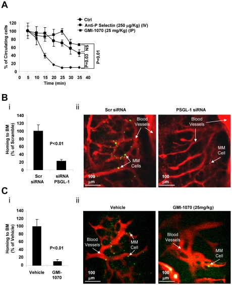
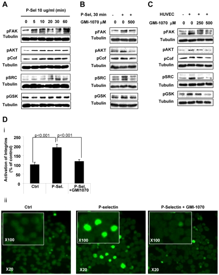
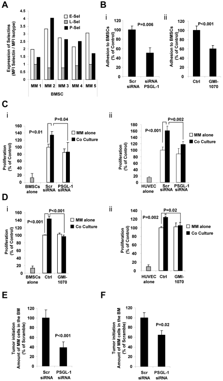
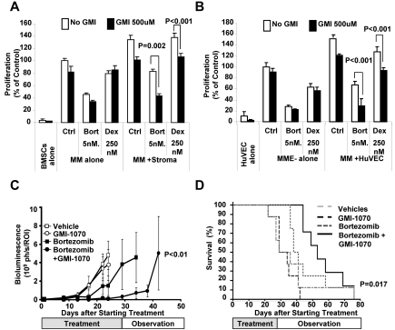
Interactions between multiple myeloma (MM) cells and the BM microenvironment play a critical role in the pathogenesis of MM and in the development of drug resistance by MM cells. Selectins are involved in extravasation and homing of leukocytes to target organs. In the present study, we focused on adhesion dynamics that involve P-selectin glycoprotein ligand-1 (PSGL-1) on MM cells and its interaction with selectins in the BM microenvironment. We show that PSGL-1 is highly expressed on MM cells and regulates the adhesion and homing of MM cells to cells in the BM microenvironment in vitro and in vivo. This interaction involves both endothelial cells and BM stromal cells. Using loss-of-function studies and the small-molecule pan-selectin inhibitor GMI-1070, we show that PSGL-1 regulates the activation of integrins and downstream signaling. We also document that this interaction regulates MM-cell proliferation in coculture with BM microenvironmental cells and the development of drug resistance. Furthermore, inhibiting this interaction with GMI-1070 enhances the sensitization of MM cells to bortezomib in vitro and in vivo. These data highlight the critical contribution of PSGL-1 to the regulation of growth, dissemination, and drug resistance in MM in the context of the BM microenvironment.


Figures

Figure 1
Figure 1
PSGL-1 is highly expressed on MM cells and regulates interaction with P-selectins. (A) Expression of PSGL-1 detected in BM biopsies from MM patients (n = 17) and healthy individuals (n = 3) using immunohistochemistry. Images are showing 20× magnification and inserts are showing 100× magnification. All MM patients presented with a higher expression of PSGL-1 compared with healthy individuals. (B) Expression of PSGL-1 evaluated on MM cell lines (MM.1S, OPM1, OPM2, LR7, RPMI.8226, U266, and H929) using flow cytometry and expressed as a ratio between the MFI of PSGL-1 and the MFI of the isotype control. (C) Gene-expression profiling of PSGL-1 from available dataset series number GSE 6477. The expression level of PSGL-1 significantly increases with MM tumor progression from MGUS to smoldering MM to newly diagnosed MM. Significant differences are observed between healthy subjects and MM patients (both smoldering MM and newly diagnosed MM) (P < .01). (D) CD138+ cells isolated from either normal BM (n = 3) or MM BM (n = 6) and MM cell lines (MM.1S, OPM1, OPM2, RPMI.8226, U266, LR7, and H929) incubated with free human Fc-chain (isotype control) or with chimeras of human-Fc chain and recombinant human E-, L-, or P-selectin (10 μg/mL) for 1 hour, followed by FITC-conjugated mouse anti–human Fc. The interaction of the selectins with MM cells was analyzed by flow cytometry and quantified as the ratio of the MFI of each selectin to the MFI of the isotype control. P- and L-selectins (but not E-selectin) interacted with MM primary cells and cell lines, whereas none of the selectins interacted with the normal plasma cells. (E) MM1s cells were transfected with either PSGL-1 siRNA or scramble siRNA. Cells were exposed to recombinant human E-, L-, and P-selectin (10 μg/mL) for 1 hour. The interaction of selectins with MM cells was analyzed by flow cytometry and quantified as ratio of MFI of each selectin to the MFI of the isotype control. Down-regulation of PSGL-1 reduced the interaction of both L- and P-selectins with MM cells. (F) MM.1S cells were treated with increasing concentrations of GMI-1070 (0, 250, and 500μM) for 1 hour and then exposed to recombinant E-, L-, and P-selectin (10 μg/mL, for 1 hour). The interaction of selectins with MM cells was analyzed by flow cytometry and quantified as a ratio of MFI of each selectin to the MFI of the isotype control. Dose-dependent inhibition of the interaction of L- and P-selectins was observed. All data represent means ± SD of triplicate experiments.
Figure 2
Figure 2
PSGL-1 regulated adhesion of MM cells to ECs. (A) Expression of E-, L-, and P-selectins evaluated on HUVECs and primary MM BM-derived ECs (n = 5) using flow cytometry and expressed as ratio between MFI of selectin to the MFI of the isotype control. ECs present with higher expression of E- and P-selectins. (B) HUVECs were transfected with siRNAs for E-, L-, or P-selectin. Scramble siRNA was used as a control (panel i) or cells were treated with anti–E-, anti–L-, or anti–P-neutralizing Abs. Isotype control Ab was used as a control (panel ii). Adhesion of MM cells on HUVECs was evaluated: significant inhibition of MM cells to HUVECs was observed in P-selectin knock-down cells (panel i) and in HUVECs treated with neutralizing Ab for P-selectin (panel ii), P < .01. All HUVECs shown in panel C were exposed to TNFα (30 U/mL for 3 hours) or to IL-4 (3 ng/mL for 24 hours) and histamine (2.25mM for 4 hours). Expression of E-, L-, and P-selectins was evaluated by flow cytometry. Induction of E- and P-selectin was observed after activation with TNFα or IL-4/histamine, respectively (panel i). Adhesion of nontreated MM cells to HUVECs was evaluated. MM cells showed increased adhesion to HUVECs with activation of P-selectin (panel ii), P < .01. (D) MM1s cells were transfected with either PSGL-1 siRNA or scramble siRNA. Adhesion of MM cells to HUVECs was evaluated: a significant inhibition of MM cells to HUVECs was observed in PSGL-1 knock-down cells (P < .05). (E) HUVECs were treated with increasing concentrations of GMI-1070 (0, 100, 250, and 500μM) for 1 hour, and adhesion of nontreated MM cells (H929, OPM1, and MM.1S) to HUVECs was evaluated: dose-dependent inhibition of MM cell adhesion to HUVECs was observed (P < .05). All data represent means ± SD of triplicate experiments. (F) MM1s cells were treated with either isotype control or PSGL-1–blocking Ab. Adhesion of MM cells to HUVECs was evaluated: significant inhibition of MM cells to HUVECs was observed in cells treated with PSGL-1–blocking Ab (P < .01).
Figure 3
Figure 3
PSGL-1 regulates transendothelial migration of MM cells. (A) HUVECs were transfected with siRNAs for E-, L-, or P-selectin. Scramble siRNA was used as a control (panel i) or cells were treated with anti–E-, anti–L-, or anti–P-neutralizing Abs. Isotype control Ab was used as a control (panel ii). Transendothelial migration of MM1s cells in response to SDF-1α (30nM) was tested. Significant inhibition of MM cell transendothelial migration was observed in HUVECS with P-selectin knockdown (panel i) and in HUVECs treated with neutralizing Ab for P-selectin (panel ii) (P < .01). (B) MM1s cells were transfected with either PSGL-1 or scramble siRNA, and transendothelial migration of MM cells in response to SDF-1α (30nM) was tested. Significant inhibition of MM cell transendothelial migration was observed in MM cells with PSGL-1 knockdown. (C) HUVECs were treated with increasing concentrations of GMI-1070 (0, 100, 250, and 500μM) for 1 hour, and transendothelial migration of nontreated MM1s cells in response to SDF-1α (30nM) was evaluated: dose-dependent inhibition of MM cell transendothelial migration was observed (P < .02). (D) MM1s cells were transfected with either PSGL-1 or scramble siRNA, and migration of MM cells (with no presence of HUVECs) in response to SDF-1α (30nM) was tested. No difference in cell migration was observed in MM cells transfected with PSGL-1 or scramble siRNA. (E) MM1s cells were treated with increasing concentrations of GMI-1070 (0, 100, 250, and 500μM) for 1 hour, and migration of MM cells (with no presence of HUVECs) in response to SDF-1α (30nM) was tested: no difference in cell migration was observed in MM cells treated with or without GMI-1070. (F) MM1s cells were treated with either isotype control or PSGL-1–blocking Ab. Transendothelial migration of MM cells was evaluated: significant inhibition of MM cells to HUVECs was observed in cells treated with PSGL-1–blocking Ab.
Figure 4
Figure 4
The interaction of PSGL-1 with P-selectin regulates extravasation and homing to the BM of MM cells in vivo. (A) Calcein-labeled MM1s cells were injected intravenously into SCID mice treated with either vehicle (Ctrl), anti–P-selectin Ab (250 μg/kg IV), or GMI-1070 (25 mg/kg IP) for 1 hour before injection of MM1s cells (n = 3 per group). The number of circulating cells was followed over time using in vivo flow cytometry. Cells were counted every 5 minutes for 40 minutes. Fluorescence signal was detected on an artery in the ear and digitized for analysis with MATLAB software. Inhibition of P-selectin using GMI-1070 or neutralizing Ab delayed the extravasation of MM cells. (B) MM1s cells were transfected with either PSGL-1 or scramble siRNA, labeled with Calcein, and injected intravenously into SCID mice (n = 3 per group), followed by IV injection of Evans blue. Homing to the BM of mice was imaged by in vivo confocal microscopy 30 minutes after injection. Inhibition of the homing of MM cells to the BM was observed with knockdown of PSGL-1, shown as an average of number of MM cells in 18 images taken from 3 different mice (P < .01; i) and in representative images of the BM (ii; green indicates MM cells; red, blood vessels). (C) Calcein-labeled MM1s cells were injected intravenously into SCID mice, which were treated with either vehicle (Ctrl) or GMI-1070 (25 mg/kg IP) for 1 hour before injection of MM1s cells (n = 3 per group), followed by IV injection of Evans blue. Homing to the BM of mice was imaged by in vivo confocal microscopy 30 minutes after injection. Inhibition of the homing of MM cells to the BM was observed in mice treated with GMI-1070, shown as an average of number of MM cells in 18 images taken from 3 different mice (P < .01; i) and in representative images of the BM (ii; green indicates MM cells; red, blood vessels).
Figure 5
Figure 5
Interaction of PSGL-1 and P-selectin regulates adhesion-related signaling and β-integrin activation in MM cells. (A) MM1s cells were treated with recombinant P-selectin 10μg/mL for different durations (0, 5, 10, 20, 30, and 60 minutes), lysed, and whole-cell lysates were subjected to Western blotting for pFAK, pAKT, pCoffilin, pSRC, and p-GSK3α/β. Increased adhesion-related signaling was observed after activation with recombinant P-selectin, with maximal activation at 30 minutes. (B) Recombinant P-selectin was incubated with or without GMI-1070 (500mM for 1 hour) and then applied to MM1s cells; nontreated MM cells were used as a control. Cells were then lysed and whole-cell lysates were subjected to Western blotting for pFAK, pAKT, pCoffilin, pSRC, and p-GSK3α/β. GMI-1070 reversed the induction of adhesion-related signaling in MM cells induced by recombinant P-selectin. (C) HUVECs were treated with or without GMI-1070 (500mM for 1 hour), nontreated MM1s cells were cocultured with the HUVECs for 1 hour, and MM1s cells not cocultured with HUVECs served as a control. MM cells were then separated from the HUVECs, lysed, and whole-cell lysates were subjected to Western blotting for pFAK, pAKT, pCoffilin, pSRC, and p-GSK3α/β. Coculture of MM cells with HUVECs induced adhesion-related signaling in MM cells that was reversed by GMI-1070. (D) Recombinant P-selectin was incubated with or without GMI-1070 (500mM for 1 hour) and then applied to MM1s cells; nontreated MM cells were used as a control. Cells were fixed and the expression of activated β1-integrin was detected using FITC-labeled Ab under immunofluorescence microscopy. Increased activation of β1-integrin was observed after activation with recombinant P-selectin, which was reversed by GMI-1070. Results are shown as an average of the percentage of cells with activated integrins, normalized to controls (P < .001; i), and in representative fluorescent images at 20× magnification and at 100× in the inset (ii).
Figure 6
Figure 6
The interaction of PSGL-1 and P-selectin regulates proliferation of MM cells induced by BMSCs and ECs in vitro and tumor initiation in vivo. (A) Expression of E-, L-, and P-selectins evaluated in primary BMSCs (n = 5) using flow cytometry and expressed as ratio between MFI of selectin to the MFI of the isotype control. BMSCs presented with higher expression of E- and P-selectins. (B) MM1s cells were transfected with either PSGL-1 siRNA or scramble siRNA. Adhesion of MM cells to BMSCs was evaluated: significant inhibition of MM cell adhesion to BMSCs was observed in PSGL-1 knockdown cells (P = .006; i). BMSCs were treated with or without GMI-1070 (500μM for 1 hour) and adhesion of nontreated MM1s cells to BMSCs was evaluated: inhibition of MM cell adhesion to BMSCs was observed in HUVECs treated with GMI-1070 (P < .001; ii). Data represent means ± SD of triplicate experiments. (C) MM1s cells were transfected with either PSGL-1 siRNA or scramble siRNA and cultured with or without BMSCs (i) and HUVECs (ii). Cell proliferation was measured at 24 hours by bromodeoxyuridine incorporation and ELISA. Coculture of MM1s with HUVECs and BMSCs increased the proliferation of MM1s cells transfected with scramble siRNA, an effect that was reversed by PSGL-1 siRNA. Data represent means ± SD of triplicate experiments. (D) HUVECs and BMSCs were treated with or without GMI-1070 and nontreated MM cells were cultured with or without BMSCs (i) and HUVECs (ii). Cell proliferation was measured at 24 hours by bromodeoxyuridine incorporation and ELISA. Coculture of MM1s with nontreated HUVECs and BMSCs increased the proliferation of MM1s cells, an effect that was reversed by GMI-1070. Data represent means ± SD of triplicate experiments. (E) MM1s cells were transfected with either PSGL-1 or scramble siRNA and injected intravenously into SCID mice (n = 4 per group); after 1 week the BM was extracted from the femurs of the mice and tumor initiation was determined as the percentage of CD138+ cells in the BM. Inhibition of tumor initiation in the BM of the mice was observed with knockdown of PSGL-1 (P < .001). (F) MM1s cells were transfected with either PSGL-1 or scramble siRNA and injected into the BM of the tibia of SCID mice; after 1 week the BM was extracted from the tibias and tumor initiation was determined as the percentage of CD138+ cells in the BM. Inhibition of tumor initiation in the BM of the mice was observed with knockdown of PSGL-1, but not to the same extent as that observed after IV injection (P = .02).
Figure 7
Figure 7
The interaction of PSGL-1 and P-selectin regulates drug resistance of MM cells induced by BMSCs and ECs in vitro and tumor progression in vivo. (A) MM1s cells (treated with vehicle, bortezomib 5nM, or dexamethasone 250nM) were cultured with or withoutBMSCs (treated with or without GMI-1070 500μM). Cell proliferation was measured at 24 hours by bromodeoxyuridine incorporation and ELISA. Coculture of MM1s with BMSCs induced resistance to both bortezomib and dexamethasone in MM1s cells which was reversed by GMI-1070. Data represent means ± SD of triplicate experiments. (B) MM1s cells (treated with vehicle, bortezomib 5nM or dexamethasone 250 nM) were cultured with or without the presence of HUVECs (treated with or without GMI-1070 500μM). Cell proliferation was measured at 24 hours by bromodeoxyuridine incorporation and ELISA. Coculture of MM1s with HUVECs induced resistance to both bortezomib and dexamethasone in MM1s cells that was reversed by GMI-1070. Data represent means ± SD of triplicate experiments. (C) Effect of inhibition of the interaction of PSGL-1 with P-selectin by GMI-1070 on the sensitivity of MM tumors to bortezomib in vivo. SCID mice (n = 10 per group) were injected with MM1s cells engineered to express luciferase and tumor growth was determined by bioluminescence imaging. Mice were divided in 4 groups: (1) the control group, which received weekly IP injection of vehicle and were implanted with pumps loaded with vehicle every 2 weeks (for 4 weeks); (2) the GMI-1070–treated group, which received weekly IP injection of vehicle and were implanted with a pump loaded with 200 μL of 150 mg/mL of GMI-1070 that was changed every 2 weeks (for 4 weeks); (3) the bortezomib-treated group, which received weekly IP injection of bortezomib 1.5 mg/kg and were implanted with a pump loaded with vehicle that was changed every 2 weeks (for 4 weeks); and (4) the combination group, which received weekly IP injection of bortezomib 1.5 mg/kg and were implanted with a pump loaded with 200 μL of 150 mg/mL GMI-1070 that was changed every 2 weeks (for 4 weeks). Tumor progression was detected using bioluminescence. Treatment with GMI-1070 alone did not affect the tumor progression, but it increased the sensitivity of MM cells to bortezomib, and decreased tumor progression was observed in the group treated with a combination of GMI-1070 and bortezomib compared with bortezomib alone (P < .01). (D) Kaplan-Meier curves of survival of groups 1 through 4 described in panel C. Increased survival was observed in mice treated with the combination of GMI-1070 and bortezomib compared with bortezomib alone, (P = .012).

Comment in

References

    1. Alsayed Y, Ngo H, Runnels J, et al. Mechanisms of regulation of CXCR4/SDF-1 (CXCL12)-dependent migration and homing in multiple myeloma. Blood. 2007;109(7):2708–2717. - PMC - PubMed
    1. Lapidot T, Dar A, Kollet O. How do stem cells find their way home? Blood. 2005;106(6):1901–1910. - PubMed
    1. Damiano JS, Cress AE, Hazlehurst LA, Shtil AA, Dalton WS. Cell adhesion mediated drug resistance (CAM-DR): role of integrins and resistance to apoptosis in human myeloma cell lines. Blood. 1999;93(5):1658–1667. - PMC - PubMed
    1. Hideshima T, Chauhan D, Hayashi T, et al. The biological sequelae of stromal cell-derived factor-1alpha in multiple myeloma. Mol Cancer Ther. 2002;1(7):539–544. - PubMed
    1. Pagnucco G, Cardinale G, Gervasi F. Targeting multiple myeloma cells and their bone marrow microenvironment. Ann N Y Acad Sci. 2004;1028:390–399. - PubMed

Publication types

MeSH terms