

Chondrosarcoma of the spine: a series of 16 cases and a review of the literature
- PMID: 22096435
- PMCID: PMC3215129
Chondrosarcoma of the spine: a series of 16 cases and a review of the literature
Abstract
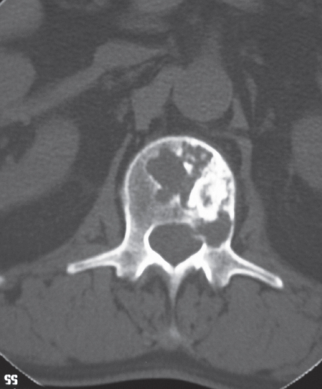
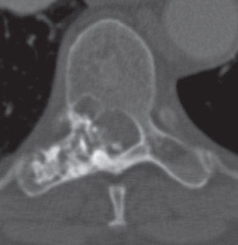
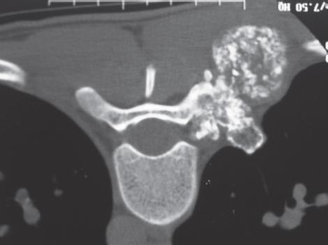
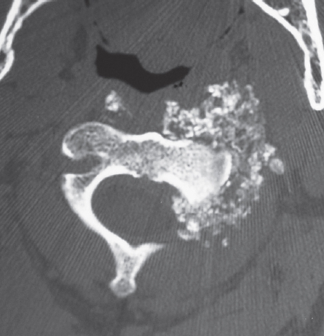
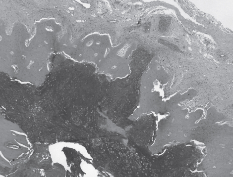
Only a few major studies of chondrosarcoma of the mobile spine have been reported. These studies have shown that spinal chondrosarcomas require complete surgical resection and are notoriously resistant to chemotherapy and radiation. We present 16 cases of chondrosarcoma of the mobile spine diagnosed at a median age of 54.5 (range 20 - 79) years. Diagnosis and treatment studies were based on both CT scans and MRI. Fifteen of our 16 patients had low-grade (grade 1-2) chondrosarcomas. All patients were treated with surgical resection. Fourteen patients had total resection while two patients had subtotal resection. The two patients who had subtotal resection died of their disease. Five of the fourteen patients who had total resection also died. The mean interval to death was 3.6 years. This study confirms that although chondrosarcomas of the spine are low grade, they are dangerous neoplasms. Even with complete resection, they have a high rate of recurrence and metastasis.
Figures

References
-
- Sundaresan N, Rosen G, Boriani S. Primary Malignant Tumors of the Spine. Orthop Clin North Am. 2009;40(1):21–36. January 1. - PubMed
-
- Tessitore E, Burkhardt K, Payer M. Primary Clear-Cell Chondrosarcoma of the Cervical Spine. J Neurosurg Spine. 2006;4(5):424. May 1. - PubMed
-
- Knoeller SM, Uhl M, Gahr N, Adler CP, Herget GW. Differential diagnosis of primary malignant bone tumors in the spine and sacrum. The radiological and clinical spectrum. Neoplasma. 2008;55(1):16–22. January 1. - PubMed
-
- Bergh P, Gunterberg B, Meis-Kindblom JM, Kindblom LG. Prognostic Factors and Outcome of Pelvic, Sacral, and Spinal Chondrosarcomas. Cancer. 2001;91(7):1201–12. April 1. - PubMed
Publication types
MeSH terms
LinkOut - more resources
Full Text Sources