. 2011;6(11):e26101.
doi: 10.1371/journal.pone.0026101. Epub 2011 Nov 9.

Toll-like receptors 2 and 4 regulate the frequency of IFNγ-producing CD4+ T-cells during pulmonary infection with Chlamydia pneumoniae

Affiliations

Toll-like receptors 2 and 4 regulate the frequency of IFNγ-producing CD4+ T-cells during pulmonary infection with Chlamydia pneumoniae

Nina Wantia et al. PLoS One. 2011.

Abstract

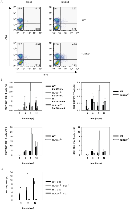
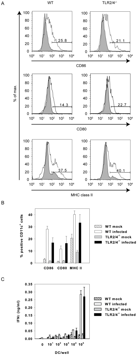
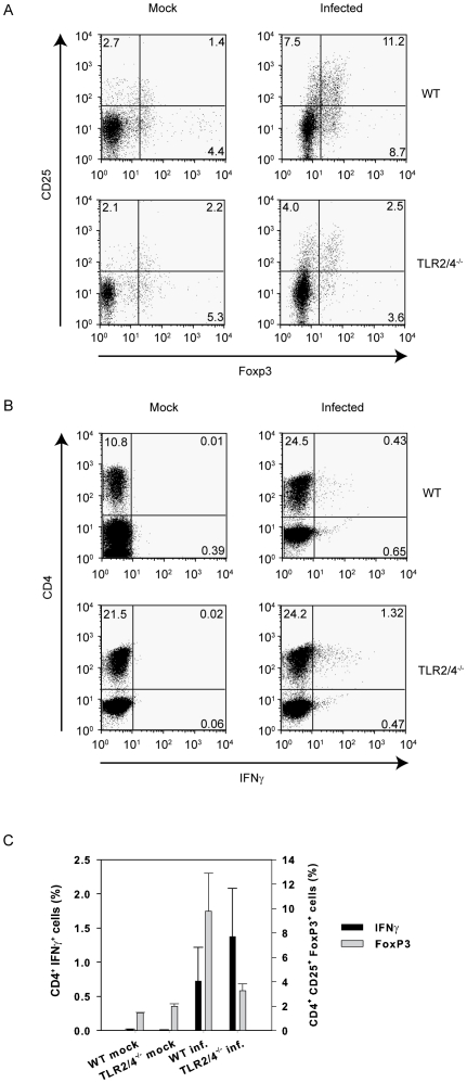
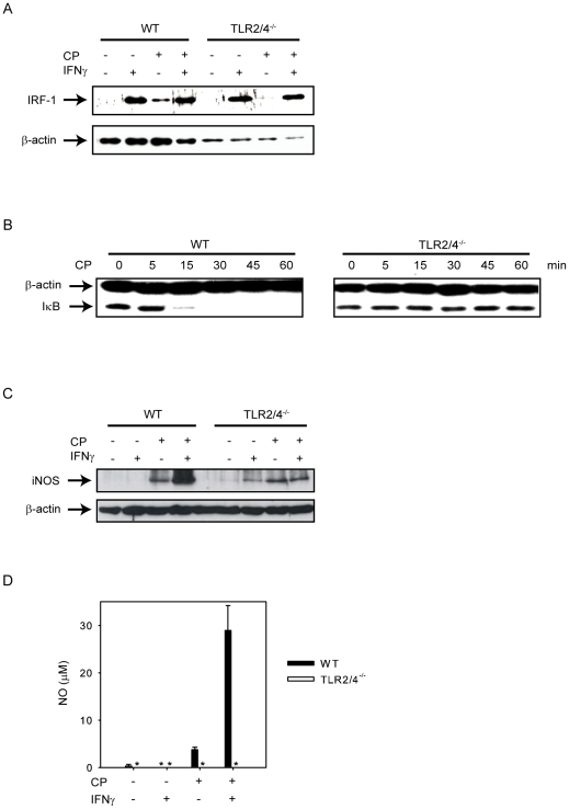
TLR2 and TLR4 are crucial for recognition of Chlamydia pneumoniae in vivo, since infected TLR2/4 double-deficient mice are unable to control the infection as evidenced by severe loss of body weight and progressive lethal pneumonia. Unexpectedly, these mice display higher pulmonary levels of the protective cytokine IFNγ than wild type mice. We show here, that antigen-specific CD4(+) T-cells are responsible for the observed IFNγ-secretion in vivo and their frequency is higher in TLR2/4 double-deficient than in wild type mice. The capacity of TLR2/4 double-deficient dendritic cells to re-stimulate CD4(+) T-cells did not differ from wild type dendritic cells. However, the frequency of CD4(+)CD25(+)Foxp3(+) T-cells was considerably higher in wild type compared to TLR2/4 double-deficient mice and was inversely related to the number of IFNγ-secreting CD4(+) effector T-cells. Despite increased IFNγ-levels, at least one IFNγ-mediated response, protective NO-secretion, could not be induced in the absence of TLR2 and 4. In summary, CD4(+)CD25(+)Foxp3(+) regulatory T-cells fail to expand in the absence of TLR2 and TLR4 during pulmonary infection with C. pneumoniae, which in turn enhances the frequency of CD4(+)IFNγ(+) effector T-cells. Failure of IFNγ to induce NO in TLR2/4 double-deficient cells represents one possible mechanism why TLR2/4 double-deficient mice are unable to control pneumonia caused by C. pneumoniae and succumb to the infection.


Conflict of interest statement

Competing Interests: The authors have declared that no competing interests exist.

Figures

Figure 1
Figure 1. TLR2 and 4 are crucial to control pneumonia-induced loss of body weight of C. pneumoniae-infected mice.
Wild type (n = 24 d0, n = 18 d6, n = 12 d9, n = 6 d12) and TLR2/4 double-deficient mice (n = 24 d0, n = 18 d6, n = 12 d9, n = 6 d12) were infected with C. pneumoniae. At the time point indicated in the graph the body weight of the mice was determined. Bars represent mean and SD of individual mice from two independent experiments. *p<0.001, ANOVA posthoc Holm-Sidak.
Figure 2
Figure 2. Lack of TLR2/4 does not impair pulmonary recruitment of CD4+ and CD8+ T-cells upon infection with C. pneumonia.
Wild type (n = 3/time point) and TLR2/4 double-deficient mice (n = 3/time point) were infected with C. pneumoniae. At the time points indicated in the graph mice were sacrificed, lungs removed, single cell suspensions were prepared and the number of cells determined. Cells were stained with mAbs specific for CD4, CD8 and CD3 as described in Materials and Methods, analyzed by flow cytometry and the number of each subpopulation was calculated. CD4+CD3+ T-cells are depicted in (A), CD8+CD3+ T-cells in (B). Error bars represent SD of three individual mice.
Figure 3
Figure 3. C. pneumoniae-specific CD4+ T cells are responsible for the enhanced IFNγ-response in TLR2/4 double-deficient mice.
(A) Pulmonary cells were prepared from wild type and TLR2/4 double-deficient mice nine days post infection with C. pneumoniae. The cells (1×10⁵ cells/well) were then re-stimulated with BMDC (1×10⁵ cells/well) which were or were not infected with C. pneumoniae (MOI = 5). After 1 h of culture Brefeldin A was added, the culture continued for another 12 h and cells were stained for CD4, CD8 and intracellular IFNγ. FACS graphs show the IFNγ response of CD4+ T-cells. (B) Pulmonary cells from C. pneumoniae-infected wild type (n = 3/time point) and TLR2/4 (n = 3/time point) double-deficient mice were prepared at different time points as indicated in the graph. Mock infected animals served as controls (time point 0). Cells were stained with mAbs specific for CD3, CD4, CD8 and IFNγ as described in Materials and Methods and analyzed by flow cytometry. Upper graphs show frequncies, lower graphs absolute numbers of IFNγ+CD4+ or CD8+ T cells. (C) Pulmonary cells were prepared from C. pneumoniae-infected wild type (n = 2/time point) of TLR2/4 double-deficient mice (n = 2 6d, n = 1 9d, n = 12d) after 6, 9, and 12 days post infection and the cells were stained for CD4, CD3 and IFNγ. Note that CD4+CD3 cells hardly contribute to IFNγ-secretion.
Figure 4
Figure 4. Enhanced extra-pulmonary IFNγ responses in C. pneumoniae-infected TLR2/4 double-deficient mice.
Lung (upper graph) and spleen cells (lower graph) were prepared nine days post infection with C. pneumoniae. Subsequently cells (1×10⁵ cells/well) were re-stimulated with BMDC (1×10⁵ cells/well) which were or were not infected with C. pneumoniae (MOI = 5) for three days. The secretion of IFNγ was determined by analyzing the culture supernatant with a commercially available ELISA-system. The graph represents the data of three independent experiments. Error bars represent SD of individual mice (n = 3 mock wild type and mock TLR2/4 double deficient mice, n = 5 infected wild type mice, n = 4 infected TLR2/4 double-deficient mice). *p = 0.016, Mann-Whitney Rank sum test.
Figure 5
Figure 5. Antigen-presentation is not impaired in TLR2/4 double-deficient BMDC.
In (A) and (B) lung cells were prepared three days post infection with C. pneumoniae. The cells were stained with antibodies specific for CD11c, CD86, CD80 or MHC class II. All events are gated on CD11c. Error bars represent SD of three individual mice. The experiment was repeated once with similar results. (C) Pulmonary cells were prepared from wild type mice infected nine days earlier with C. pneumoniae. The cells (1×10⁵ cells/well) were re-stimulated with titrated amounts of BMDC from wild type or TLR2/4 double-deficient mice as indicated which were or were not infected with C. pneumoniae (MOI = 5). The IFNγ-content of the culture supernatant was analyzed by ELISA after three days of culture. Error bars represent SD of three replicate cultures. The experiment was repeated twice with similar results.
Figure 6
Figure 6. Inverse relation in vivo between the percentage of CD4+CD25+Foxp3+ T-cells and the percentage of CD4+IFNγ+ T-cells.
Pulmonary cells were prepared from wild type and TLR2/4 double-deficient mice nine days post infection with C. pneumoniae. In (A) the expression of CD25 on the cell membrane and intracellular Foxp3 was analyzed. All events were gated on CD4. (B) shows the expression of intracellular IFNγ by CD4+ T-cells ex vivo without re-stimulation. (C) demonstrates the relationship between CD4+Foxp3+ and CD4+IFNγ+ T-cells. Error bars represent SD of three individual mice. The experiment was repeated twice with similar results.
Figure 7
Figure 7. IFNγ is severely impaired to increase iNOS expression and fails to release NO in C. pneumoniae-infected TLR2/4 double-deficient BMDMs.
(A) Wild type or TLR2/4 double-deficient BMDMs (7.5×10⁵ cells/well) were left untreated, or stimulated with IFNγ (10 ng/ml) for 24 h, or were infected with C. pneumoniae (MOI = 10, for 48 h), or were infected with C. pneumoniae for 48 h and treated with IFNγ 24 h post infection. IRF-1 was detected by Western blot, detection of β-actin was used as loading control. (B) Wild type or TLR2/4 double-deficient BMDMs (7.5×10⁵ cells/well) were or were not infected with C. pneumoniae (MOI = 10) for the time periods indicated in the graph. IκB was detected by Western blot, detection of β-actin was used as loading control. (C) Wild type or TLR2/4 double-deficient BMDMs were treated as described in (A). iNOS was determined by Western blot, detection of β-actin was used as loading control. (D) Wild type or TLR2/4 double-deficient BMDMs (7.5×10⁵ cells/well) were left untreated, or stimulated with IFNγ (10 ng/ml) for 48 h, or were infected with C. pneumoniae (MOI = 10, for 72 h), or were infected with C. pneumoniae for 72 h and treated with IFNγ 48 h post infection. Subsequently, NO-levels were determined in the culture supernatant. Error bars represent SD of four or in the case of untreated samples of two individual cultures. *not detectable. The experiment was repeated once with similar results.

References

    1. Kuo CC, Jackson LA, Campbell LA, Grayston JT. Chlamydia pneumoniae (TWAR). ClinMicrobiolRev. 1995;8:451–461. - PMC - PubMed
    1. Rodriguez N, Fend F, Jennen L, Schiemann M, Wantia N, et al. Polymorphonuclear Neutrophils Improve Replication of Chlamydia pneumoniae In Vivo upon MyD88-Dependent Attraction. J Immunol. 2005;174:4836–4844. - PubMed
    1. Summersgill JT, Sahney NN, Gaydos CA, Quinn TC, Ramirez JA. Inhibition of Chlamydia pneumoniae growth in HEp-2 cells pretreated with gamma interferon and tumor necrosis factor alpha. InfectImmun. 1995;63:2801–2803. - PMC - PubMed
    1. Rottenberg ME, Gigliotti-Rothfuchs A, Wigzell H. The role of IFN-gamma in the outcome of chlamydial infection. CurrOpinImmunol. 2002;14:444–451. - PubMed
    1. Rothfuchs AG, Gigliotti D, Palmblad K, Andersson U, Wigzell H, et al. IFN-alpha beta-dependent, IFN-gamma secretion by bone marrow-derived macrophages controls an intracellular bacterial infection. JImmunol. 2001;167:6453–6461. - PubMed

Publication types

MeSH terms