. 2011;6(11):e26976.
doi: 10.1371/journal.pone.0026976. Epub 2011 Nov 9.

Identification of residues in the heme domain of soluble guanylyl cyclase that are important for basal and stimulated catalytic activity

Affiliations

Identification of residues in the heme domain of soluble guanylyl cyclase that are important for basal and stimulated catalytic activity

Padmamalini Baskaran et al. PLoS One. 2011.

Abstract

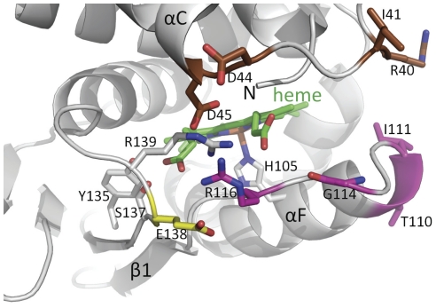
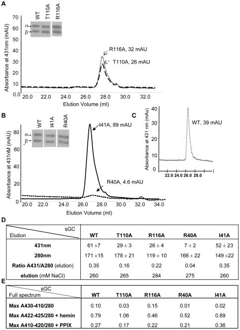
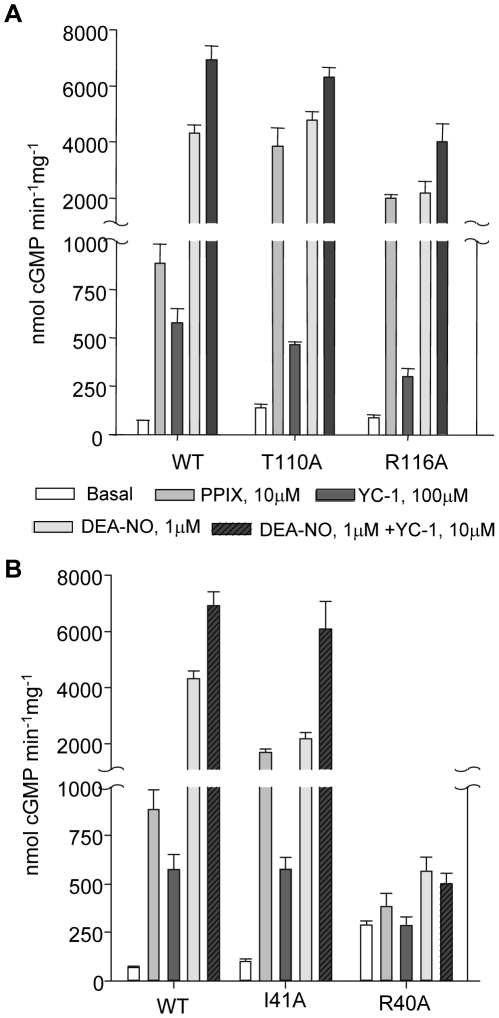
Nitric oxide signals through activation of soluble guanylyl cyclase (sGC), a heme-containing heterodimer. NO binds to the heme domain located in the N-terminal part of the β subunit of sGC resulting in increased production of cGMP in the catalytic domain located at the C-terminal part of sGC. Little is known about the mechanism by which the NO signaling is propagated from the receptor domain (heme domain) to the effector domain (catalytic domain), in particular events subsequent to the breakage of the bond between the heme iron and Histidine 105 (H105) of the β subunit. Our modeling of the heme-binding domain as well as previous homologous heme domain structures in different states point to two regions that could be critical for propagation of the NO activation signal. Structure-based mutational analysis of these regions revealed that residues T110 and R116 in the αF helix-β1 strand, and residues I41 and R40 in the αB-αC loop mediate propagation of activation between the heme domain and the catalytic domain. Biochemical analysis of these heme mutants allows refinement of the map of the residues that are critical for heme stability and propagation of the NO/YC-1 activation signal in sGC.


Conflict of interest statement

Competing Interests: The authors have declared that no competing interests exist.

Figures

Figure 1
Figure 1. Close-up view of the heme-binding region of a homology model of the heme domain of sGC.
The heme (green) and its H105 ligand are depicted. Residues targeted for mutagenesis in the region between the αB-αC helices are colored brown, targeted residues in the αF-β1 strand region are shown in magenta and the flanking E138 residue in yellow. The αC helix, β1 strand, and the αF helix containing the heme-liganding residue H105 are labeled. Residues Y135, S137, and R139 as part of the YxSxR heme binding motif are shown in grey with the residues labeled.
Figure 2
Figure 2. Purification of Wild Type and mutants.
2A–B: representative spectra of purification of mutants at 431 nm, the expected absorbance for the heme-containing enzyme. All mutants had similar levels of expression as assessed by immunoblot with antibodies against α and β subunit of sGC after electrophoresis of ∼0.5 µg of semi-purified protein on a 7.5% Tris-HCl gel (insets). 2C: representative elution profile of purification of WT. 2D: Table of purification with ratio of heme-containing sGC (431 nm) over protein total concentration (280 nm) from the elution, measured as described in Material and Methods ; values are expressed in absorbance unit (mAU). Elution as a function of salt concentration is indicated (NaCl, mM). Measurement at 393 nm to estimate oxidized heme-containing sGC was not significantly different between WT and mutants (not shown). 2E: Table of full spectrum values. UV-vis was recorded as described in Material and Methods (see also Figure S3) and the ratio of maximum absorption for WT and mutants between 410 and 430 nm over maximum absorption at 280 nm was calculated in the absence or presence of hemin (5 µM) and PPIX (5 µM) after heme reduction with 5 mM DTT. Max: Maxima. Each mutant was purified at least three times (±S.E.M.) and WT was purified six times (± S.E.M.). UV-vis collection of the various Soret bands for WT and mutants are shown in Figure S3 and Figure S4.
Figure 3
Figure 3. Basal and stimulated activities of purified WT and mutants.
3A: In the αF-β1 region, basal activity of α1/β1T110A as well as PPIX activation of α1/β1T110A and α1/β1R116A were significantly higher than WT. 3B: In the αB-αC loop, mutant α1/β1I41A exhibits a significantly higher response to PPIX and significantly lower response to DEA-NO compared to WT, yet sensitivity to DEA-NO +YC-1 was comparable to WT. α1/β1R40A lost the ability to respond to any activators. Measurements of activities were done at least three times on 50 ng of two to three independently semi-purified WT and mutants with each assay done in duplicate under each condition. Results are expressed in nmol cGMP. mg−1.min−1 ± S.E.M. Values and fold stimulation are provided in Table S1.
Figure 4
Figure 4. Response of WT, α1/β1T110A and α1/β1I41A to DEA-NO, PPIX and both.
4A: DEA-NO concentration-response curve of purified WT and mutants. cGMP production was measured as a function of increasing concentrations of DEA-NO. 4B: PPIX concentration-response curves of purified WT and mutants. cGMP production was measured as a function of increasing concentrations of PPIX. 4C: Comparison of WT and mutants' response to DEA-NO (1 µM), PPIX (10 µM) and DEA-NO (1 µM)+PPIX (10 µM). Results are expressed in nmol cGMP. mg−1.min−1 ± S.E.M. Typically, 50 ng of semi-purified enzyme was used per assay tube. Each experiment, carried out in duplicate, was repeated independently two or three times with two separate purified preparations. For the concentration-response curves, EC50 and maximal velocity were determined using Sigmaplot software (version 11.0).

References

    1. Ignarro LJ, Cirino G, Casini A, Napoli C. Nitric oxide as a signaling molecule in the vascular system: an overview. J Cardiovasc Pharmacol. 1999;34:879–886. - PubMed
    1. Garthwaite J. New insight into the functioning of nitric oxide-receptive guanylyl cyclase: physiological and pharmacological implications. Mol Cell Biochem. 2010;334:221–232. - PubMed
    1. Derbyshire ER, Marletta MA. Biochemistry of soluble guanylate cyclase. Handb Exp Pharmacol. 2009:17–31. - PubMed
    1. Mergia E, Koesling D, Friebe A. Genetic mouse models of the NO receptor ‘soluble’ guanylyl cyclases. Handb Exp Pharmacol. 2009:33–46. - PubMed
    1. Ma X, Sayed N, Baskaran P, Beuve A, van den Akker F. PAS-mediated dimerization of soluble guanylyl cyclase revealed by signal transduction histidine kinase domain crystal structure. J Biol Chem. 2008;283:1167–1178. - PMC - PubMed

Publication types