Clinical Trial
. 2011;6(11):e27351.
doi: 10.1371/journal.pone.0027351. Epub 2011 Nov 9.

IL-2 stimulated but not unstimulated NK cells induce selective disappearance of peripheral blood cells: concomitant results to a phase I/II study

Affiliations
Clinical Trial

IL-2 stimulated but not unstimulated NK cells induce selective disappearance of peripheral blood cells: concomitant results to a phase I/II study

Claudia Brehm et al. PLoS One. 2011.

Abstract

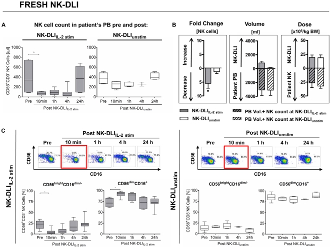
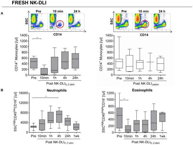
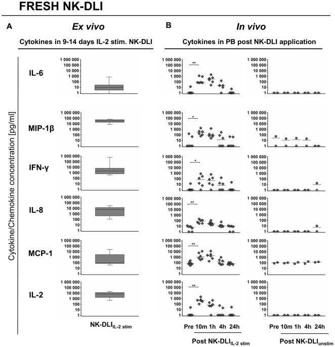
In an ongoing clinical phase I/II study, 16 pediatric patients suffering from high risk leukemia/tumors received highly purified donor natural killer (NK) cell immunotherapy (NK-DLI) at day (+3) +40 and +100 post haploidentical stem cell transplantation. However, literature about the influence of NK-DLI on recipient's immune system is scarce. Here we present concomitant results of a noninvasive in vivo monitoring approach of recipient's peripheral blood (PB) cells after transfer of either unstimulated (NK-DLI(unstim)) or IL-2 (1000 U/ml, 9-14 days) activated NK cells (NK-DLI(IL-2 stim)) along with their ex vivo secreted cytokine/chemokines. We performed phenotypical and functional characterizations of the NK-DLIs, detailed flow cytometric analyses of various PB cells and comprehensive cytokine/chemokine arrays before and after NK-DLI. Patients of both groups were comparable with regard to remission status, immune reconstitution, donor chimerism, KIR mismatching, stem cell and NK-DLI dose. Only after NK-DLI(IL-2 stim) was a rapid, almost complete loss of CD56(bright)CD16(dim/-) immune regulatory and CD56(dim)CD16(+) cytotoxic NK cells, monocytes, dendritic cells and eosinophils from PB circulation seen 10 min after infusion, while neutrophils significantly increased. The reduction of NK cells was due to both, a decrease in patients' own CD69(-) NCR(low)CD62L(+) NK cells as well as to a diminishing of the transferred cells from the NK-DLI(IL-2 stim) with the CD56(bright)CD16(+/-)CD69(+)NCR(high)CD62L(-) phenotype. All cell counts recovered within the next 24 h. Transfer of NK-DLI(IL-2 stim) translated into significantly increased levels of various cytokines/chemokines (i.e. IFN-γ, IL-6, MIP-1β) in patients' PB. Those remained stable for at least 1 h, presumably leading to endothelial activation, leukocyte adhesion and/or extravasation. In contrast, NK-DLI(unstim) did not cause any of the observed effects. In conclusion, we assume that the adoptive transfer of NK-DLI(IL-2 stim) under the influence of ex vivo and in vivo secreted cytokines/chemokines may promote NK cell trafficking and therefore might enhance efficacy of immunotherapy.


Conflict of interest statement

Competing Interests: The authors have declared that no competing interests exist.

Figures

Figure 1
Figure 1. NK-DLIIL-2 stim but not NK-DLIunstim led to a considerable disappearance of NK cells from PB.
A) Absolute number of NK cells in the PB of the patients was significantly reduced 10 min post NK-DLIIL-2 stim (grey) in contrast to NK-DLIunstim (white), which showed minimal influence only. 24 h after NK-DLIIL-2 stim absolute number of NK cells recovered to the level before DLI. Only freshly applied NK-DLIs infused around day +40 post SCT are shown and were used for statistical calculations (n = 7 NK-DLIIL-2 stim, n = 6 NK-DLIunstim). Box and whiskers plots show minimum, lower quartile, median, upper quartile and maximum of all measured data. For the 4 h level post NK-DLIunstim two values were available, only. p<0.05 indicated as *. B) Mean and SEM of all freshly applied NK-DLIIL-2 stim (grey) and NK-DLIunstim (white) applied around day +40 post SCT. Striped bars indicate estimated patient's PB volume and peripheral NK cell count at the time point of NK-DLI infusion. Left graph shows a mean 5.5-fold reduction of absolute NK cell count in the PB as early as 10 min post NK-DLIIL-2 stim compared to a 1.2-fold decrease for NK-DLIunstim. Middle graph shows relation of NK-DLI volume to total blood volume of patients (NK-DLIIL-2 stim volume: 825 ml±249, compared to PB volume: 3060 ml±789; NK-DLIunstim volume: 128 ml±19, compared to PB volume 3093 ml±749). DLI volume did not artificially lead to the reduced absolute NK cell count, since NK-DLIIL-2 stim volumes made up maximally around ¼ of patient's PB volume. Right graph shows mean NK-DLI cell dose ×106/kg BW (NK-DLIIL-2 stim 19.2±4.5; NK-DLIunstim 18.8±5.1) in relation to patient's PB NK cells newly reconstituted post haplo-SCT around d +40 prior to NK-DLI (NK-DLIIL-2 stim 26.0±8.5; NK-DLIunstim 31.9±4.0). Applied NK dose compromised about 80% of PB NK cells, illustrating the high dose of administered NK-DLI. C) Density plots (CD56 vs. CD16) and box and whiskers plots show a significant change in the distribution of the cytotoxic CD56dimCD16+ and immune regulatory CD56brightCD16dim/− NK cell subsets. This was due to an absolute reduction of the CD56brightCD16dim/− NK cell subpopulation in the PB 10 min after freshly applied NK-DLIIL-2 stim applications infused around d +40 (left; n = 7). This could not be shown after NK-DLIunstim (right; n = 6). Plots are gated on CD56+CD3 NK cells. For the 4 h level post NK-DLIunstim 2 values were available, only. p<0.05 indicated as *.
Figure 2
Figure 2. Influence of NK-DLIs on monocyte and granulocyte distribution in patient's PB.
A) Significant reduction of absolute CD14+ monocyte count could be demonstrated in patient's PB 10 min after application of fresh NK-DLIIL-2 stim (n = 7), while recovering to normal values within the next 24 h. This was not seen after NK-DLIunstim (n = 6). Density plots show side scatter (SSC) vs. CD14 gated on CD45+ leukocytes. For the 4 h level post NK-DLIunstim 2 values were available, only. p<0.05 indicated as *. B) We observed a distinct influence of NK-DLIIL-2 stim on neutrophil and eosinophil granulocyte count. A massive increase of neutrophils (SSChighCD45lowCD16+) with its peak at 4 h was combined with a significant reduction of eosinophil granulocytes (SSChighCD45brightCD16) in the PB after fresh NK-DLIIL-2 stim (n = 7). This was not observed after unstimulated NK cell applications (data not shown). p<0.05 indicated as *.
Figure 3
Figure 3. In vivo monitoring of patient's cytokine/chemokine plasma levels following NK-DLI.
A) Box and whiskers plots show the respective cytokine/chemokine concentration present in the NK-DLIIL-2 stim product (n = 12) immediately prior to infusion. The nine to 14 days ex vivo IL-2 stimulation of highly purified donor NK cells led to the secretion of high amounts of various chemokines i.e. IL-8 and MIP-1β, as well as the pro-inflammatory cytokine IFN-γ. Whereas, the median concentration of the pro-inflammatory cytokine IL-6 was <10 pg/ml. The indicated IL-2 concentration was due to addition during expansion procedure (see Material and Methods). Y-axis shows cytokine/chemokine concentration, range 1–1.000.000 pg/ml. B) Cytokine analyses of cryopreserved plasma samples collected before (pre) and 10 min, 1 h, 4 h and 24 h after fresh NK-DLIIL-2 stim (⧫, n = 6). Significant increases of in vivo cytokine/chemokine concentration of IL-2, IL-6, IL-8, IFN-γ, MCP-1 and MIP-1β in patient's plasma following NK-DLIIL-2 stim were observed. Levels peaked after 10 min to 1 h post infusion, remained enhanced over a period of 4 h and returned to base level within the next 24 h. In contrast, no increase in cytokine/chemokine PB concentration following NK-DLIunstim was seen (•, n = 2). p<0.05 and <0.01 indicated as * and **.

References

    1. Passweg JR, Koehl U, Uharek L, Meyer-Monard S, Tichelli A. Natural-killer-cell-based treatment in haematopoietic stem-cell transplantation. Best Pract Res Clin Haematol. 2006;19:811–824. - PubMed
    1. Passweg JR, Stern M, Koehl U, Uharek L, Tichelli A. Use of natural killer cells in hematopoetic stem cell transplantation. Bone Marrow Transplant. 2005;35:637–643. - PubMed
    1. Rubnitz JE, Inaba H, Ribeiro RC, Pounds S, Rooney B, et al. NKAML: a pilot study to determine the safety and feasibility of haploidentical natural killer cell transplantation in childhood acute myeloid leukemia. J Clin Oncol. 2010;28:955–959. - PMC - PubMed
    1. Lanier LL. NK cell recognition. Annu Rev Immunol. 2005;23:225–274. - PubMed
    1. Moretta A, Bottino C, Vitale M, Pende D, Cantoni C, et al. Activating receptors and coreceptors involved in human natural killer cell-mediated cytolysis. Annu Rev Immunol. 2001;19:197–223. - PubMed

Publication types