. 2011;6(11):e27510.
doi: 10.1371/journal.pone.0027510. Epub 2011 Nov 11.

Atypical scrapie isolates involve a uniform prion species with a complex molecular signature

Affiliations

Atypical scrapie isolates involve a uniform prion species with a complex molecular signature

Dorothea R Götte et al. PLoS One. 2011.

Abstract

The pathobiology of atypical scrapie, a prion disease affecting sheep and goats, is still poorly understood. In a previous study, we demonstrated that atypical scrapie affecting small ruminants in Switzerland differs in the neuroanatomical distribution of the pathological prion protein (PrP(d)). To investigate whether these differences depend on host-related vs. pathogen-related factors, we transmitted atypical scrapie to transgenic mice over-expressing the ovine prion protein (tg338). The clinical, neuropathological, and molecular phenotype of tg338 mice is similar between mice carrying the Swiss atypical scrapie isolates and the Nor98, an atypical scrapie isolate from Norway. Together with published data, our results suggest that atypical scrapie is caused by a uniform type of prion, and that the observed phenotypic differences in small ruminants are likely host-dependant. Strikingly, by using a refined SDS-PAGE technique, we established that the prominent proteinase K-resistant prion protein fragment in atypical scrapie consists of two separate, unglycosylated peptides with molecular masses of roughly 5 and 8 kDa. These findings show similarities to those for other prion diseases in animals and humans, and lay the groundwork for future comparative research.


Conflict of interest statement

Competing Interests: The authors have declared that no competing interests exist.

Figures

Figure 1
Figure 1. Survival of tg338 mice inoculated with different TSE isolates.
Mice infected with Swiss atypical scrapie isolates (red) showed survival times similar to those infected with Nor98 (black), but these survival times were clearly shorter compared to the classical scrapie group (blue) and sham-inoculated mice (grey).
Figure 2
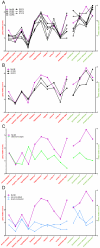
Figure 2. Lesion profiles in scrapie-infected tg338 mice.
Lesions were scored according to the protocol established by Fraser and Dickinson in ten grey matter areas (left y-axis, scale 0 to 5) and in four white matter areas (right y-axis, scale 0 to 3). All depicted values represent means of scores in mice inoculated with the same isolate: atypical sheep scrapie (A), atypical goat scrapie (B), classical scrapie (C) as well as sham-inculated and non-inoculated mice (D). To facilitate cross-comparison, Nor98 scores are included in each graph.
Figure 3
Figure 3. Representative spongiform pathology in tg338 mouse brains.
(A) Vacuolar lesions are evident in the hypothalamus, hippocampus, corpus callosum, and cerebral peduncle of TSE-inoculated and control mice. Classical scrapie-inoculated mice exhibit plaques in various brain structures such as the hippocampus (star). (B) Variability of spongiform lesions in the corpus callosum between mice inoculated with atypical scrapie isolates. (C) Cerebellum of a non-inoculated mouse showing mild (left) and a sham-inoculated mouse showing severe (right) vacuolar lesions. All microphotographs are 40X magnifications of hematoxylin and eosin-stained mouse brain sections.
Figure 4
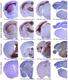
Figure 4. Neuroanatomical PrPd distribution in mice inoculated with different scrapie isolates.
Microphotographs of representative mouse brain sections stained for PrPd by immunohistochemistry using antibody SAF84 are presented. For details on the anatomical structures, see Fig S1.
Figure 5
Figure 5. Localization and size of plaque-like PrP deposits in atypical and classical scrapie.
In atypical scrapie-inoculated mice, relatively small plaques were observed in the neuropil (40X magnification), while the plaques in the classical scrapie-inoculated mice were larger, coalescing and always associated to the leptomeninges and the ependyma (20X magnification). PrPd immunoreactivity was detected using the mAb SAF84. Neuroanatomical structures: septal nuclei (se), caudate putamen (pu), lateral ventricle (lv) and thalamus (th).
Figure 6
Figure 6. Molecular PrPres typing of atypical scrapie affected tg338 mice.
Western blots for PrPres of representative brain extracts following SDS-PAGE using (A) 16.5% tris-glycine and (B-F) 4–20% tris-glycine gradient gels. Immunochemical detection was performed with antibodies P4, Sha31 or 9A2 as indicated. In some instances (D, E), samples were deglycosylated with PNGaseF. (F) Atypical scrapie PrPres banding pattern following sample pretreatment with different concentrations of proteinase K (PK, in µg/ml), compared to the PK digestion protocol of the Bio-Rad TeSeE confirmatory Western blot (BR). Identification codes for the atypical scrapie isolates are shown at the top. Controls: cl, classical scrapie affected tg338 mouse; non-inoc, non-inoculated tg 338 mouse; neg s, TSE negative sheep. Molecular mass standards in kDa and loaded tissue equivalents are indicated.
Figure 7
Figure 7. Molecular PrPres typing of atypical scrapie-affected sheep and goats.
SDS-PAGE was performed using 4–20% gradient gels and blots were probed with mAb P4 (A) and Sha31 (B). Samples of a classical scrapie-affected sheep (cl) and a TSE-negative sheep (neg) served as controls. Molecular mass standards and tissue equivalents are indicated.

References

    1. Prusiner SB. Molecular biology of prion diseases. Science. 1991;252:1515–1522. - PubMed
    1. Griffith JS. Self-replication and scrapie. Nature. 1967;215:1043–1044. - PubMed
    1. Hope J, Morton LJ, Farquhar CF, Multhaup G, Beyreuther K, et al. The major polypeptide of scrapie-associated fibrils (SAF) has the same size, charge distribution and N-terminal protein sequence as predicted for the normal brain protein (PrP). EMBO J. 1986;5:2591–2597. - PMC - PubMed
    1. Schneider K, Fangerau H, Michaelsen B, Raab WH. The early history of the transmissible spongiform encephalopathies exemplified by scrapie. Brain Res Bull. 2008;77:343–355. - PubMed
    1. Foster JD, Hope J, Fraser H. Transmission of bovine spongiform encephalopathy to sheep and goats. Vet Rec. 1993;133:339–341. - PubMed

Publication types