

Proliferative tumor doubling times of prostatic carcinoma
- PMID: 22096656
- PMCID: PMC3196972
- DOI: 10.1155/2011/301850
Proliferative tumor doubling times of prostatic carcinoma
Abstract
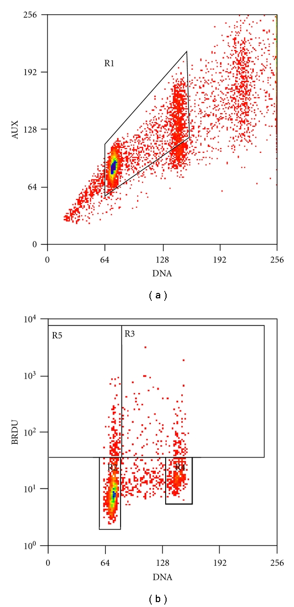
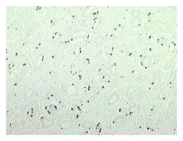
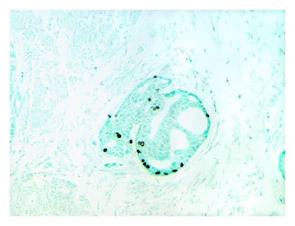
Prostate cancer (PCa) has a variable biology ranging from latent cancer to extremely aggressive tumors. Proliferative activities of cancers may indicate their biological potential. A flow cytometric assay to calculate maximum proliferative doubling times (T(max)) of PCa in radical prostatectomy specimens after preoperative in vivo bromodeoxyuridine (BrdU) infusion is presented. Only 4/17 specimens had tumors large enough for flow cytometric analysis. The T(max) of tumors was similar and ranged from 0.6 to 3.6 months. Tumors had calculated doubling times 2- to 25-fold faster than their matched normal tissue. Variations in labeling index and T(max) were observed within a tumor as well as between different Gleason grades. The observed PSA doubling times (PSA-DT) ranged from 18.4 to 32.0 months, considerably slower than the corresponding T(max) of tumors involved. While lack of data for apoptotic rates is a limitation, apparent biological differences between latent versus aggressive PCa may be attributable to variations in apoptotic rates of these tumors rather than their cell proliferative rates.
Figures
Similar articles
-
More advantages in detecting bone and soft tissue metastases from prostate cancer using 18F-PSMA PET/CT.Hell J Nucl Med. 2019 Jan-Apr;22(1):6-9. doi: 10.1967/s002449910952. Epub 2019 Mar 7. Hell J Nucl Med. 2019. PMID: 30843003
-
Preoperative serum prostate specific antigen levels between 2 and 22 ng./ml. correlate poorly with post-radical prostatectomy cancer morphology: prostate specific antigen cure rates appear constant between 2 and 9 ng./ml.J Urol. 2002 Jan;167(1):103-11. J Urol. 2002. PMID: 11743285
-
Pretreatment prostate specific antigen doubling times: use in patients before radical prostatectomy.J Urol. 1997 Nov;158(5):1876-8; discussion 1878-9. doi: 10.1016/s0022-5347(01)64154-5. J Urol. 1997. PMID: 9334621 Clinical Trial.
-
Best practices recommendations in the application of immunohistochemistry in the prostate: report from the International Society of Urologic Pathology consensus conference.Am J Surg Pathol. 2014 Aug;38(8):e6-e19. doi: 10.1097/PAS.0000000000000238. Am J Surg Pathol. 2014. PMID: 25029122
-
Radical prostatectomy for impalpable prostate cancer: the Johns Hopkins experience with tumors found on transurethral resection (stages T1A and T1B) and on needle biopsy (stage T1C).J Urol. 1994 Nov;152(5 Pt 2):1721-9. doi: 10.1016/s0022-5347(17)32370-4. J Urol. 1994. PMID: 7523719 Review.
Cited by
-
Systematic identification of functionally relevant risk alleles to stratify aggressive versus indolent prostate cancer.Oncotarget. 2018 Feb 5;9(16):12812-12824. doi: 10.18632/oncotarget.24400. eCollection 2018 Feb 27. Oncotarget. 2018. PMID: 29560112 Free PMC article.
-
Molecular natural history of breast cancer: Leveraging transcriptomics to predict breast cancer progression and aggressiveness.Cancer Med. 2020 May;9(10):3551-3562. doi: 10.1002/cam4.2996. Epub 2020 Mar 23. Cancer Med. 2020. PMID: 32207233 Free PMC article.
-
An integrative model of prostate cancer interaction with the bone microenvironment.Math Biosci. 2017 Dec;294:1-14. doi: 10.1016/j.mbs.2017.09.005. Epub 2017 Sep 14. Math Biosci. 2017. PMID: 28919575 Free PMC article.
-
Cancer recurrence times from a branching process model.PLoS Comput Biol. 2019 Nov 21;15(11):e1007423. doi: 10.1371/journal.pcbi.1007423. eCollection 2019 Nov. PLoS Comput Biol. 2019. PMID: 31751332 Free PMC article.
-
Didox (3,4-dihydroxybenzohydroxamic acid) suppresses IgE-mediated mast cell activation through attenuation of NFκB and AP-1 transcription.Cell Immunol. 2017 Dec;322:41-48. doi: 10.1016/j.cellimm.2017.09.008. Epub 2017 Sep 21. Cell Immunol. 2017. PMID: 28964543 Free PMC article.
References
-
- Jemal A, Siegel R, Xu J, Ward E. Cancer statistics, 2010. CA Cancer Journal for Clinicians. 2010;60(5):277–300. - PubMed
-
- Miller GJ, Cygan JM. Morphology of prostate cancer: the effects of multifocality on histological grade, tumor volume and capsule penetration. Journal of Urology. 1994;152(5):1709–1713. - PubMed
-
- Sakr WA, Grignon DJ, Crissman JD, et al. High grade prostatic intraepithelial neoplasia (HGPIN) and prostatic adenocarcinoma between the ages of 20–69: an autopsy study of 249 cases. In Vivo. 1994;8(3):439–443. - PubMed
-
- Crawford ED, Hirano D, Werahera PN, et al. Computer modeling of prostate biopsy: tumor size and location—not clinical significance—determine cancer detection. Journal of Urology. 1998;159(4):1260–1264. - PubMed
-
- Hirano D, Werahera PN, Crawford ED, Lucia MS, DeAntoni EP, Miller GJ. Morphological analysis and classification of latent prostate cancer using a 3-dimensional computer algorithm: Analysis of tumor volume, grade, tumor doubling time and life expectancy. Journal of Urology. 1998;159(4):1265–1269. - PubMed
Grants and funding
LinkOut - more resources
Full Text Sources
Research Materials
Miscellaneous
