. 2012 Feb;31(2):110-8.
doi: 10.5732/cjc.011.10327. Epub 2011 Nov 18.

Overexpression of P-glycoprotein induces acquired resistance to imatinib in chronic myelogenous leukemia cells

Affiliations

Overexpression of P-glycoprotein induces acquired resistance to imatinib in chronic myelogenous leukemia cells

Xing-Xiang Peng et al. Chin J Cancer. 2012 Feb.

Abstract

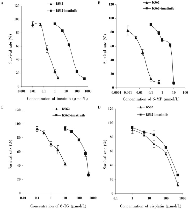
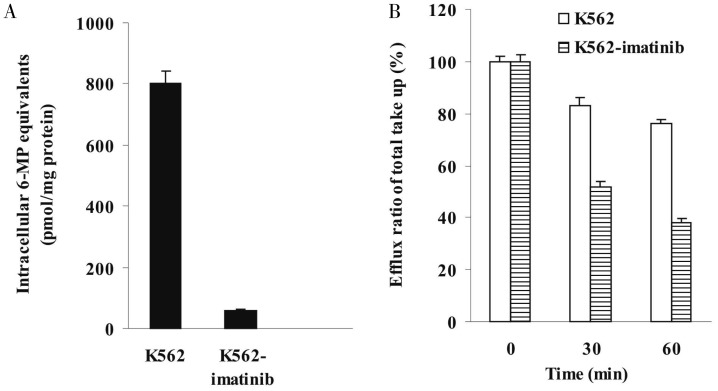
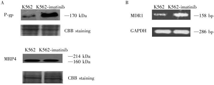
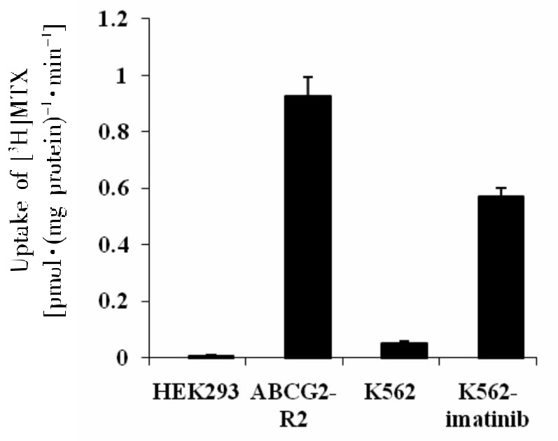
Imatinib, a breakpoint cluster region (BCR)-Abelson murine leukemia(ABL) tyrosine kinase inhibitor (TKI), has revolutionized the treatment of chronic myelogenous leukemia (CML). However, development of multidrug resistance(MDR) limits the use of imatinib. In the present study, we aimed to investigate the mechanisms of cellular resistance to imatinib in CML. Therefore, we established an imatinib-resistant human CML cell line(K562-imatinib) through a stepwise selection process. While characterizing the phenotype of these cells, we found that K562-imatinib cells were 124.6-fold more resistant to imatinib than parental K562 cells. In addition, these cells were cross-resistant to second- and third-generation BCR-ABL TKIs. Western blot analysis and reverse transcription-polymerase chain reaction(RT-PCR) demonstrated that P-glycoprotein(P-gp) and MDR1 mRNA levels were increased in K562-imatinib cells. In addition, accumulation of [14C]6-mercaptopurine (6-MP) was decreased, whereas the ATP-dependent efflux of [14C]6-MP and [3H]methotrexate transport were increased in K562-imatinib cells. These data suggest that the overexpression of P-gp may play a crucial role in acquired resistance to imatinib in CML K562-imatinib cells.


Figures

Figure 1.
Figure 1.. Sensitivity of K562 cells (▴) and K562-imatinib cells (▪) to imatinib (A), 6-mercaptopurine (6-MP) (B), 6-thioguanine (6-TG) (C), and cisplatin (D).
The data were analyzed using the MTT cytotoxicity assay as described in “Materials and Methods” section. Data points are presented as means ± standard deviation (SD) of at least three separate experiments.
Figure 2.
Figure 2.. Time-dependent accumulation and efflux of [14C]6-mercaptopurine (6-MP) and/or its metabolites in K562 and K562-imatinib cells.
A, accumulation of [14C]6-MP and/or its metabolites at 60 min in K562 and K562-imatinib cells in complete medium. Values are presented as mean ± SD of three separate experiments. Data were analyzed by unpaired Student's t-test. Accumulation of [14C]6-MP and/or its metabolites at 60 min in K562-imatinib cells decreased significantly compared with that in K562 cells (P < 0.001). B, time course of efflux of [14C]6-MP and/or its metabolites in K562 and K562-imatinib cells after various efflux times. Values are presented as mean + SD of three separate experiments. The percentages of retained 6-MP equivalents after 30 and 60 min efflux are indicated in K562 and K562-imatinib cells relative to that at 0 min. Data were analyzed by unpaired Student's t-test. After 30 and 60 min efflux, the retained 6-MP equivalents in K562-imatinib cells decreased significantly compared with that in K562 cells (P < 0.001).
Figure 3.
Figure 3.. Expression of P-glycoprotein (P-gp) and multidrug resistance protein 4 (MRP4) in K562 and K562-imatinib cells.
A, Western blot analysis of P-gp and MRP4 in membrane vesicle preparations from K562 and K562-imatinib cells. Membrane vesicles were prepared as described in Materials and Methods. Protein was resolved by SDS-PAGE on 4%-12% gel and electrotransferred to nitrocellulose membranes. Mobilities of the molecular mass markers are indicated in kilodaltons. The bottom panel is a section of an identical gel stained with coomassie brilliant blue (CBB) to demonstrate approximately equal loading. B, reverse transcription-polymerase chain reaction (RT-PCR) analysis of mRNA expression levels of MDR1 in K562 and K562-imatinib cells.
Figure 4.
Figure 4.. Uptake of [3H] methotrexate (MTX) into inside-out membrane vesicles derived from K562 and K562-imatinib cells.
Values are presented as mean ± SD of three separate experiments. Membrane vesicles prepared from HEK293/pcDNA and HEK293/ABCG2-R2 are used as negative and positive controls, respectively. Data were analyzed by unpaired Student's t-test. The ATP-dependent uptake of [3H]MTX in K562-imatinib cells increased significantly compared with K562 cells (P < 0.001).

References

    1. Rowley JD. A new consistent chromosomal abnormality in chronic myelogenous leukaemia identified by quinacrine fluorescence and Giemsa staining. Nature. 1973;243:290–293. - PubMed
    1. Goldman JM, Melo JV. Chronic myeloid leukemia-advances in biology and new approaches to treatment. N Engl J Med. 2003;349:1451–1464. - PubMed
    1. Hehlmann R, Hochhus A, Baccarani M. Chronic myeloid leukemia. Lancet. 2007;370:342–350. - PubMed
    1. Roskoski R., Jr STI-571: an anticancer protein-tyrosine kinase inhibitor. Biochem Biophys Res Commun. 2003;309:709–717. - PubMed
    1. Walz C, Sattler M. Novel targeted therapies to overcome imatinib mesylate resistance in chronic myeloid leukemia (CML) Crit Rev Oncol Hematol. 2006;57:145–164. - PubMed

Publication types

MeSH terms