. 2011 Nov 18;44(4):647-59.
doi: 10.1016/j.molcel.2011.08.043.

Telomere protection by TPP1/POT1 requires tethering to TIN2

Affiliations

Telomere protection by TPP1/POT1 requires tethering to TIN2

Kaori K Takai et al. Mol Cell. .

Erratum in

Abstract

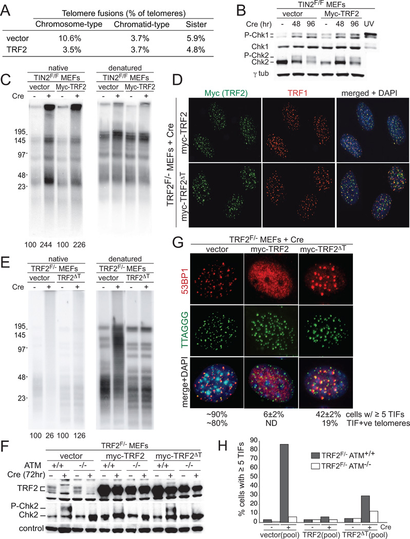
To prevent ATR activation, telomeres deploy the single-stranded DNA binding activity of TPP1/POT1a. POT1a blocks the binding of RPA to telomeres, suggesting that ATR is repressed through RPA exclusion. However, comparison of the DNA binding affinities and abundance of TPP1/POT1a and RPA indicates that TPP1/POT1a by itself is unlikely to exclude RPA. We therefore analyzed the central shelterin protein TIN2, which links TPP1/POT1a (and POT1b) to TRF1 and TRF2 on the double-stranded telomeric DNA. Upon TIN2 deletion, telomeres lost TPP1/POT1a, accumulated RPA, elicited an ATR signal, and showed all other phenotypes of POT1a/b deletion. TIN2 also affected the TRF2-dependent repression of ATM kinase signaling but not to TRF2-mediated inhibition of telomere fusions. Thus, while TIN2 has a minor contribution to the repression of ATM by TRF2, its major role is to stabilize TPP1/POT1a on the ss telomeric DNA, thereby allowing effective exclusion of RPA and repression of ATR signaling.


Figures

Figure 1
Figure 1. Abundance and DNA binding features of POT1, TPP1/POT1a and RPA
(A) and (B) Quantitative immunoblotting for RPA32, POT1a, and POT1b in whole cell lysates and comparison to recombinant standards (Fig. S1A). Right: abundance of human RPA and mouse POT1a/b based on data from three experiments as shown on the left. (C) Gel-shift reactions with the indicated probes and increasing amounts (0.16–40 nM) of human POT1 or RPA. (D) Binding of mouse TPP1/POT1a and human RPA to Tel34. Protein amounts as in (C). (E) Summary of the apparent Kd of human RPA, human POT1, and mouse TPP1/POT1a derived from gel-shift experiments as shown in (C) and (D) (average values from three experiments represented and standard deviations). Kd values were derived from mathematical curve fitting (GraphPad Prism) of PhosphorImager data on RPA and POT1 analyzed in parallel.
Figure 2
Figure 2. Conditional deletion of TIN2
(A) Schematic showing the Tinf2 genomic locus, the targeting construct, and the alleles generated. Black triangles, LoxP sites with BglI/BglII sites; black boxes, Frt sites; Neo, PGK-Neo gene; DTA, MCI-DTA; black bars, probes for genomic blotting; asterisk, TRF1 interaction motif in exon 6. Right: PCR genotyping of TIN2+/+ and TIN2F/F MEFs after introduction of Cre. (B) Loss of telomeric TIN2 signals from TIN2F/F MEFs treated with pWZL-Cre (92 h). IF for TIN2 (Ab 1447, red) at telomeres detected by FISH (green). (C) Telomeric DNA ChIP for shelterin proteins in TIN2F/F MEFs with or without Cre treatment (92 h). ChIP signals were normalized to the input and the background (PI) was subtracted. Numbers below represent the average decrease in these values after deletion of TIN2 (two experiments). (D) TIN2 deletion diminishes the telomeric IF signals for TRF1 and TRF2. Method as in (B). (E) Effect of TIN2 deletion (H&R-Cre) on soluble and chromatin-bound shelterin proteins. Equal cell equivalents of the whole cell lysate (WC), cytoplasmic proteins (CP), nucleoplasmic proteins (NP), and the chromatin-bound fraction (CB) were analyzed. α-tubulin is cytoplasmic. (F) Immunoblotting for Myc-POT1a and -TPP1 in TIN2F/F MEFs infected with pLPC-puro-Myc-TPP1 or pWZL-Hygro-Myc-POT1a and treated with Cre (92 h) after drug selection. (G) Telomeric ChIP for Myc-POT1a and -TPP1 before and after TIN2 deletion. ChIP assay with TIN2 Ab (1447) and myc Ab as in (F). Input: 20% of the input DNA. (H) Quantification of the ChIP signals in (G) after normalization to input and subtraction of background (PI). (I) Telomeric localization of Myc-TPP1 and -POT1a. IF for myc (red) and telomeric FISH (green) at 92 hr post-Cre.
Figure 3
Figure 3. Excess ss telomeric DNA, endoreduplication, and telomere fusions in TIN2 KO
(A) In-gel hybridization assay for ss telomeric DNA after TIN2 deletion. Left: TelC signals under the native condition. Right: same gel re-hybridized after in situ denaturation of the DNA. Overhang signals (left) were normalized to the total telomeric signals (right) and compared to TIN2F/F MEFs without Cre. Two independent experiments are shown. (B) FACS analysis for DNA content (propidium iodide) of TIN2F/F and TIN2+/+ MEFs after pWZL-Cre infection (day 8). Both MEFs contained tetraploid cells prior to deletion of TIN2. (C) Diplochromosomes in Cre-treated TIN2F/F MEFs. DNA stained with DAPI (blue) and telomeric FISH (green). (D) Telomere clustering in a polyploid TIN2-deficient cell. Staining for 53BP1 IF (red) and telomeric FISH (green). DNA is stained with DAPI (blue). The enlarged image illustrates clustered telomeres. (E) CO-FISH illustrating examples of telomere fusions and T-SCEs in metaphases of TIN2-deficient cells. Red: leading-end telomeres; green: lagging-end telomeres; blue: DAPI DNA stain. (F) Summary of telomere phenotypes induced by TIN2 deletion determined by CO-FISH as shown in (E). Values are averages of 3–4 independent experiments (1000–2000 telomeres/experiment) and SDs. Sister fusions were scored on long arm telomeres in metaphase spreads with separated chromosome arms.
Figure 4
Figure 4. TIN2 loss induces ATM and ATR signaling
(A) TIFs detected by IF-FISH in MEFs of the indicated genotypes. MEFs were fixed at 92 hr after H&R Cre and processed as in Fig. 3D. (B) Immunoblots of phospho-Chk1, total Chk1, and Chk2 in MEFs of the indicated genotypes at 48 and 92 hr after H&R Cre. UV treated (30 min after 25 J/m2) TIN2F/F MEFs serve as a control. (C) Quantification of the effect of ATM and ATR deletion on TIFs induced by TIN2 deletion. TIN2F/F (top), TIN2F/FATRF/F (middle), and TIN2F/FATM−/− MEFs (bottom) were scored for 53BP1 TIFs per nucleus (n>100) after H&R-Cre (92 h). Averages of 3 independent experiments and SDs. P values from one-way analysis of variance (ANOVA) and Bonferroni’s Multiple Comparison Test.
Figure 5
Figure 5. RPA at telomeres after TIN2 deletion
(A) Co-localization of RPA with telomeres in TIN2F/F MEFs infected with Cre (92 h). RPA32 IF (red) and telomeric DNA FISH (green). Arrowheads: RPA signals at telomeres. Below: quantification of cells with 5 or more telomeric RPA signals as averages from 3 experiments (n≥100 nuclei) and standard errors. Asterisks: micronuclei. The cells used also expressed Myc-RPA32 but the myc-tag was not used for IF. (B) Model for RPA exclusion through TIN2-tethered TPP1/POT1. POT1 is shown to have a greater on-rate (arrow) due to its TPP1 connection to the TIN2/TRF1/TRF2 complex on the duplex telomeric DNA. Although only the most terminal shelterin complex is depicted, POT1 in shelterin distal from the telomere terminus may well be physically close to ss DNA due to higher order structure of the telomeric DNA. Tethered POT1 may also prevent RPA binding to the ss telomeric DNA in the D loop when telomeres are in the t-loop configuration (not shown).
Figure 6
Figure 6. TRF2 requires TIN2 for repression of ATM but not NHEJ
(A) Overexpression of TRF2 reduces chromosome-type fusions in TIN2 KO cells. Telomere fusions were scored as in Figs. 3E and F. (B) Immunoblots for Chk1 and Chk2 phosphorylation. Cells as in (A) and processing as in Fig. 4B. (C) No effect of TRF2 overexpression on telomeric overhang in TIN2 KO cells. Cells as in (A) and processing and quantification as in Fig. 3A. (D) Telomeric localization of the TRF2ΔT mutant. TRF2F/−MEFs were infected with the indicated TRF2 alleles, selected, cloned, and treated with Cre. TRF2 was detected with the Myc Ab in combination with IF for TRF1. (E) Repression of telomere fusions by TRF2ΔT. Cells as in D were analyzed at 120 hr post-Cre as in Fig. 3A. (F) Chk2 phosphorylation in TRF2ΔT cells. TRF2F/−ATM+/+ and TRF2F/−ATM−/− cells were infected with the indicated retroviruses, selected, and treated with Cre as indicated. Immunblotting for Chk2 and TRF2 at 72 hr post-Cre. Loading control: non-specific band in the Chk2 blot. (G) TIFs in TRF2ΔT cells. The indicated clonal lines (as in (D)) were treated with Cre (96 h) and analyzed for TIFs as in Fig. 4A. Note fewer but larger 53BP1 TIFs in TRF2ΔT cells. (H) Quantification of the TIF response in the indicated cells at 96 hr after Cre. Pools of TRF2F/− ATM+/+ and TRF2F/−ATM−/− infected as indicated were used for TIF assays as in Fig. 4A.

References

    1. Abreu E, Aritonovska E, Reichenbach P, Cristofari G, Culp B, Terns RM, Lingner J, Terns MP. TIN2-tethered TPP1 recruits human telomerase to telomeres in vivo. Mol Cell Biol. 2010;30:2971–2982. - PMC - PubMed
    1. Arnoult N, Saintome C, Ourliac-Garnier I, Riou JF, Londono-Vallejo A. Human POT1 is required for efficient telomere C-rich strand replication in the absence of WRN. Genes Dev. 2009;23:2915–2924. - PMC - PubMed
    1. Ball HL, Ehrhardt MR, Mordes DA, Glick GG, Chazin WJ, Cortez D. Function of a conserved checkpoint recruitment domain in ATRIP proteins. Mol Cell Biol. 2007;27:3367–3377. - PMC - PubMed
    1. Ball HL, Myers JS, Cortez D. ATRIP binding to replication protein A-single-stranded DNA promotes ATR-ATRIP localization but is dispensable for Chk1 phosphorylation. Mol Biol Cell. 2005;16:2372–2381. - PMC - PubMed
    1. Baumann P, Price C. Pot1 and telomere maintenance. FEBS Lett. 2010;584:3779–3784. - PMC - PubMed

Publication types

MeSH terms