Comparative Study
. 2011 Nov 17;72(4):587-601.
doi: 10.1016/j.neuron.2011.08.029.

Recruitment of endophilin to clathrin-coated pit necks is required for efficient vesicle uncoating after fission

Affiliations
Comparative Study

Recruitment of endophilin to clathrin-coated pit necks is required for efficient vesicle uncoating after fission

Ira Milosevic et al. Neuron. .

Abstract

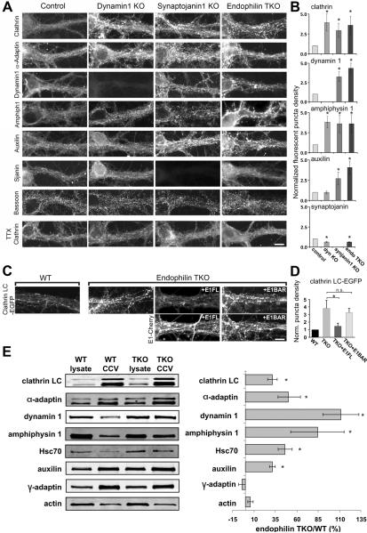
Endophilin is a membrane-binding protein with curvature-generating and -sensing properties that participates in clathrin-dependent endocytosis of synaptic vesicle membranes. Endophilin also binds the GTPase dynamin and the phosphoinositide phosphatase synaptojanin and is thought to coordinate constriction of coated pits with membrane fission (via dynamin) and subsequent uncoating (via synaptojanin). We show that although synaptojanin is recruited by endophilin at bud necks before fission, the knockout of all three mouse endophilins results in the accumulation of clathrin-coated vesicles, but not of clathrin-coated pits, at synapses. The absence of endophilin impairs but does not abolish synaptic transmission and results in perinatal lethality, whereas partial endophilin absence causes severe neurological defects, including epilepsy and neurodegeneration. Our data support a model in which endophilin recruitment to coated pit necks, because of its curvature-sensing properties, primes vesicle buds for subsequent uncoating after membrane fission, without being critically required for the fission reaction itself.


Figures

Figure 1
Figure 1. The recruitment of synaptojanin 1-145 at endocytic pits is dynamin independent, but requires endophilin
Control fibroblasts and fibroblasts that lack expression of dynamin 1 and 2 (dynamin KO) were transfected with fluorescent proteins and examined by spinning disk confocal microscopy. (A) and (B) Endophilin 2-Ruby and EGFP-synaptojanin 1 precisely colocalize in both control and dynamin KO cells (magnified in the inset). The few fluorescent spots in control cells represent very late pits (Perera et al., 2006) while the numerous puncta in dynamin KO cells represent arrested CCPs. Bar=10γm. (C) Both C-terminally EGFP-tagged full length (FL) endophilin 2 and endophilin 2 BAR domain (1–247) are recruited to the arrested CCPs in dynamin KO cells also expressing mRFP-clathrin LC. Bar=5γm (D) Model of endophilin and synaptojanin localization at the necks of the CCPs in cells with and without dynamin. (E–G) Dynamin DKO cells were transfected with synaptojanin 1-145-EGFP and clathrin LC-mRFP after siRNA-dependent KD of endophilin 2 (the major isoform in fibroblasts). (E) The synaptojanin 1–145 puncta visible in control siRNA treated cells (reflecting protein on the tubular necks of the pits) are drastically reduced after endophilin 2 KD, whose effectiveness is demonstrated by the western blot of (F). 96.0%±1.5% of the synaptojanin spots were directly adjacent to clathrin puncta (n=194 synaptojanin spots from 5 cells). Bar=5γm. (G) Quantification of the synaptojanin puncta density in the two conditions (8 cells/condition, p=0.0002).
Figure 2
Figure 2. Absence of endophilin causes perinatal lethality
(A) Endophilin 1,2 double KO (DKO) mouse and control littermate heterozygous for endophilin 1 and 2 at P18. Note the smaller size of the DKO mouse. (B) Endophilin triple KO (TKO) newborn mouse and control littermate WT for endophilin 2. Note absence of milk (arrows) in the stomach of the TKO. (C) Survival curves of endophilin TKOs, endophilin 1,2 DKOs and control mice derived from the same litters. (D) Weight at birth and growth curves of endophilin mutant mice. Note the early arrest of growth of endophilin 1,2 DKOs, but the steady growth of control littermates. Mean±SEM of mice from at least 14 litters. (E) Immunoblot analysis of brain homogenates (left) and GST pull-downs (right) from WT and endophilin KO mice with anti-endophilin isoform-specific antibodies and anti-pan-endophilin antibody. Endophilin 1 and 3 have the same electrophoretic motility (arrow), while endophilin 2 has a slower motility (arrowhead). The relevant endophilins are absent in the corresponding KO material. Note (left panel) that the absence of endophilin 1 has a greater impact than the absence of endophilin 3 on the intensity of the pan-endophilin band (arrow), indicating the greater abundance of endophilin 1 in brain. There is no difference in the expression of the remaining endophilins in the corresponding KOs. Right panel: blots of GST pull-downs from WT, DKO and TKO brain extracts using synaptojanin 1's PRD domain as a bait. (F) Histogram showing levels of synaptic proteins, as detected by quantitative immunoblotting, in brain extracts of TKO newborn mice normalized to WT. The TKO extracts show reduced levels of many SV proteins. Bars=mean±SEM (N≥4, *p<0.05, t-test).
Figure 3
Figure 3. Synaptic transmission is impaired but not abolished in endophilin TKO neurons
(A) Reduced frequency of spontaneous mEPSCs in TKO neurons (*p=0.007, t-test). The amplitude is also slightly reduced (*p=0.007, t-test). (B) The amplitudes of EPSCs in response to a single action potential (AP) are smaller in the TKO (N=20) than in control (N=19) neurons (*p=0.0027, two-tail wilcoson test). (C) Synaptic depression in response to 30 APs delivered at low (1 Hz) and high (10 Hz) frequency. Left panels show examples of individual recordings (in the 1Hz recordings, AP traces are superimposed). Right panels, which show control (N=19) and TKO (N=20) responses normalized to the first EPSC, demonstrate a difference only at the 20 Hz stimulation. (D) Decline of EPSC peak amplitude in response to a longer stimulation (300 AP delivered at 20 Hz; upper panels) and subsequent recovery upon interruption of the stimulus as detected by a 0.2 Hz test stimuli (lower panels). Depression occurred faster and recovered slower in TKO (N=17) relative to WT (N=17) neurons. EPSCs were normalized to the EPSC peak amplitude of the train.
Figure 4
Figure 4. Compensatory endocytic recovery is slower in endophilin TKO neurons
(A–B) The average time-course of the endocytic recovery after a 10 Hz 300 AP stimulus, as measured by synaptopHluorin (N=14 for WT, 13 for TKO) and vGLUT1-pHluorin (N=16 for both WT and TKO) is slower in TKO than in WT. Arrows mark the start of the stimuli. Values shown are mean±SEM. (C–D) Overexpression of full length endophilin 1-mRFP rescues the slower endocytic recovery of synaptopHluorin and vGLUT1-pHluorin signals, respectively (green traces). In contrast, an endophilin BAR construct (aa1-290)-mRFP shows only a partial rescue of the endocytic defect, which is more prominent during the late phase of the recovery (blue traces). The green and blue traces from the rescue experiments are superimposed on the recoveries shown in (A) and (B) to allow a direct comparison of the kinetics of recovery. (E) Average t1/2 recovery times for the conditions shown in (A–D). *p<0.05, t-test, error bars=SEM. (F) The delayed recovery from the increase of pHluorin fluorescence is not due to a defect in reacidification of newly endocytosed vesicles. Neurons expressing vGLUT1-pHluorin were briefly exposed to pulses of extracellular acid solution (pH 5.5) at the times indicated. In both WT (N=8) and TKO (N=8), the fluorescence quenched consistently to the same levels before and after the stimulus, arguing against a lag in the acidification of a newly internalized vesicle pool.
Figure 5
Figure 5. Increased number of clathrin-coated vesicles, but not clathrin-coated pits at endophilin DKO and TKO synapses
(A–B) EM ultrastructure of control (WT) and endophilin TKO synapses from cortical neuronal cultures (DIV21). SVs occupy nearly the entire control nerve terminal, while in the TKO synapse, the few SVs (outlined by dashed white line) are surrounded by numerous CCVs, which are sparsely packed and embedded in a dense cytomatrix. Bars=200 nm. (C–E) Endophilin TKO, synaptojanin 1 KO and dynamin 1 KO synapses, respectively. In all three synapses, a cluster of densely packed SVs (white dashed line) is surrounded by abundant coated vesicular profiles, but only in dynamin 1 KO synapses examples of such structures with elongated necks in the plane of the section can be seen. Bars=250 nm. Insets: higher magnification of clathrin-coated structures. Bars=50 nm. (F–I) Morphometric analysis of SVs, CCVs and CCPs in control and endophilin mutant synapses. The ratio of CCVs to total (CCVs+SVs) vesicles is shown in I. Each circle represents one synapse. (J–L) 3D models of control, endophilin DKO and TKO synapses derived from the reconstruction of 300 nm-thick tomograms showing SVs (blue), CCVs (green) and CCPs (yellow). Green and red lines show plasma membrane and the postsynaptic density, respectively. Bars=200 nm. (M–N) Synapses from the same brain stem region in control and DKO mice at P9. Note the only few SVs anchored to the active zone (arrowheads) and the numerous CCVs (arrows and inset) in the DKO synapse. Bars=200 nm, inset=100 nm.
Figure 6
Figure 6. Redistribution of endocytic proteins at endophilin TKO synapses
(A) Immunofluorescence of the proteins indicated at left in cortical neuronal cultures (DIV 18–24) from control, dynamin 1 KO, synaptojanin 1 KO and endophilin TKO mice. Clathrin, α–adaptin and amphiphysin 1 are predominantly diffuse in control neurons, but clustered in all three mutant genotypes. Dynamin 1 and auxilin are clustered in synaptojanin and endophilin mutant synapses, but auxilin is not clustered in dynamin 1 KO. Synaptojanin is slightly more diffuse in the absence of endophilin. The punctate Bassoon immunofluorescence is similar in all genotypes. Following treatment with TTX (1 μM, 14–18 h) to silence neuronal activity, clathrin was no longer clustered. Bar=15 μm. (B) Quantification of the clustering of immunoreactivity. The y-axis represents the fold increase of fluorescence puncta in mutant synapses normalized to controls. *p<0.05, t-test. Bars=mean±SEM. (C) Fluorescence analysis of EGFP-clathrin LC in WT and endophilin TKO neurons (DIV 18–24). Clathrin is predominantly diffuse in control neurons, but clustered in endophilin TKOs. Clustering was rescued by expression of endophilin 1 FL-Cherry, but not of the endophilin 1 BAR-Cherry construct. (D) Quantification of the clustering of the clathrin fluorescence in the four conditions shown in (C). The y-axis represents the fold increase of fluorescence puncta in mutant synapses normalized to WT. Bars=mean±SEM, n.s. not significant, *p<.05, t-test. (E) Left: Western blot analysis of starting lysates and CCV enriched fractions obtained from WT and endophilin TKO primary cultures (DIV 21). Right: Levels of proteins in the CCV enriched fractions from endophilin TKO cultures normalized to the WT values. Bars=mean±SEM, *p<0.05, t-test.
Figure 7
Figure 7. Neurodegeneration in endophilin 1, 2 DKO mice (P18)
(A) H&E stained cerebellar cortex of control (E1+/−E2+/−E3+/+) and endophilin DKO mice. Unstained vacuolar structures, suggesting a form of spongiform neurodegeneration, are present in DKO granule cell layer. Bars=100 μm. (B) EM of the DKO granule cell layer showing a vacuolar structure filled with membranous debris. Bars=1 μm. (C) EM of mossy fiber synapses in control and DKO cerebella. Note in the DKO the low number of SVs and the presence of sparsely packed CCVs (higher magnification in the inset). Bars=500 nm, inset 50 nm. (D) Morphology of climbing fibers (top row), as revealed by immunofluorescence for vGLUT2, in control and DKO mice. Sections were counterstained for the IP3 receptor, a Purkinje cell marker (middle row). Note in the overlay image that climbing fibers in the DKOs are swollen and do not extend beyond the most proximal portion of Purkinje cell dendrites. Bars=20 μm. (E) EM of climbing fiber synapses. Note the low number of SVs and the presence of sparsely packed CCVs (higher magnification in the inset) in the DKOs. Bars=250 nm, inset 50 nm.
Figure 8
Figure 8. Putative model of clathrin-coated vesicle fission and uncoating at synapses
Assembly and early maturation of endocytic CCPs is independent of endophilin. Endophilin is recruited only to the neck of late stage pits. The dynamin-endophilin interaction may regulate dynamin function, but it is dispensable for dynamin recruitment and for fission. In contrast, the synaptojanin-endophilin interaction is critically important for the fate of the vesicle after fission. Loss of PI(4,5)P2 on the bud may start before fission and be restricted to the bud due to the presence of a collar comprising endophilin, other BAR proteins and dynamin (see Discussion). Auxilin recruitment and uncoating are triggered only after fission.

Comment in

References

    1. Antonny B. Membrane deformation by protein coats. Curr Opin Cell Biol. 2006;18:386–394. - PubMed
    1. Bai J, Hu Z, Dittman JS, Pym EC, Kaplan JM. Endophilin functions as a membrane-bending molecule and is delivered to endocytic zones by exocytosis. Cell. 2010;143:430–441. - PMC - PubMed
    1. Blondeau F, Ritter B, Allaire PD, Wasiak S, Girard M, Hussain NK, Angers A, Legendre-Guillemin V, Roy L, Boismenu D, et al. Tandem MS analysis of brain clathrin-coated vesicles reveals their critical involvement in synaptic vesicle recycling. Proc Natl Acad Sci U S A. 2004;101:3833–3838. - PMC - PubMed
    1. Chang-Ileto B, Frere SG, Chan RB, Voronov SV, Roux A, Di Paolo G. Synaptojanin 1-mediated PI(4,5)P2 hydrolysis is modulated by membrane curvature and facilitates membrane fission. Dev Cell. 2011;20:206–218. - PMC - PubMed
    1. Cremona O, Di Paolo G, Wenk MR, Luthi A, Kim WT, Takei K, Daniell L, Nemoto Y, Shears SB, Flavell RA, et al. Essential role of phosphoinositide metabolism in synaptic vesicle recycling. Cell. 1999;99:179–188. - PubMed

Publication types

MeSH terms

Substances