

Molecular imaging of nuclear factor-κB in bladder as a primary regulator of inflammatory response
- PMID: 22099998
- PMCID: PMC3714865
- DOI: 10.1016/j.juro.2011.09.007
Molecular imaging of nuclear factor-κB in bladder as a primary regulator of inflammatory response
Abstract
Purpose: Nuclear factor-κB activation is implicated in chronic inflammatory disorders and it is a key regulator of genes involved in the response to infection, inflammation and stress. Interstitial cystitis and painful bladder syndrome are common inflammatory disorders of the bladder characterized by frequent urination and bladder pain. The role of nuclear factor-κB activation in bladder inflammation is not well defined.
Materials and methods: Female transgenic nuclear factor-κB-luciferase Tag mice (The Jackson Laboratory, Bar Harbor, Maine) were used to perform serial, noninvasive in vivo and ex vivo molecular imaging of nuclear factor-κB activation in the whole body after administering arsenic trioxide (5 mg/kg), lipopolysaccharide (2 mg/kg) or cyclophosphamide (Sigma®) (200 mg/kg) to initiate acute transient bladder inflammation. Pretreatment with dexamethasone (Sigma) (10 mg/kg) was used to modulate cyclophosphamide induced nuclear factor-κB dependent luminescence in vivo.
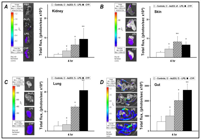
Results: Treatment of nuclear factor-κB-luciferase Tag mice with chemicals increased luminescence in a time and organ specific manner in vivo and ex vivo. The highest levels of bladder nuclear factor-κB dependent luminescence were observed 4 hours after cyclophosphamide administration. Pretreatment with dexamethasone 1 hour before cyclophosphamide injection significantly down-regulated cyclophosphamide induced bladder nuclear factor-κB dependent luminescence, ameliorated the grossly evident pathological features of acute inflammation and decreased cellular immunostaining for nuclear factor-κB in the bladder.
Conclusions: Nuclear factor-κB activity may have an important role in the pathophysiology of bladder inflammation. Nuclear factor-κB-luciferase mice can serve as a useful model in which to screen potential candidate drugs for cystitis associated with aberrant nuclear factor-κB activity. Such screening may significantly aid the development of therapeutic strategies to manage inflammatory bladder disorders.
Copyright © 2012 American Urological Association Education and Research, Inc. Published by Elsevier Inc. All rights reserved.
Figures


Similar articles
-
Sesquiterpene lactone parthenolide ameliorates bladder inflammation and bladder overactivity in cyclophosphamide induced rat cystitis model by inhibiting nuclear factor-kappaB phosphorylation.J Urol. 2009 May;181(5):2339-48. doi: 10.1016/j.juro.2009.01.015. Epub 2009 Mar 19. J Urol. 2009. PMID: 19303104
-
Nuclear factor kappa B mediates lipopolysaccharide-induced inflammation in the urinary bladder.J Urol. 2000 Mar;163(3):993-8. J Urol. 2000. PMID: 10688037
-
Molecular imaging of NF-kappaB in prostate tissue after systemic administration of IL-1 beta.Prostate. 2008 Jan 1;68(1):34-41. doi: 10.1002/pros.20666. Prostate. 2008. PMID: 18004768
-
Molecular imaging of the transcription factor NF-kappaB, a primary regulator of stress response.Mutat Res. 2004 Jul 13;551(1-2):199-211. doi: 10.1016/j.mrfmmm.2004.02.024. Mutat Res. 2004. PMID: 15225593 Review.
-
Neural upregulation in interstitial cystitis.Urology. 2007 Apr;69(4 Suppl):24-33. doi: 10.1016/j.urology.2006.08.1108. Urology. 2007. PMID: 17462476 Review.
Cited by
-
Transgenic Animal Models to Visualize Cancer-Related Cellular Processes by Bioluminescence Imaging.Front Pharmacol. 2019 Mar 15;10:235. doi: 10.3389/fphar.2019.00235. eCollection 2019. Front Pharmacol. 2019. PMID: 30930779 Free PMC article. Review.
-
Cranberry, but not D-mannose and ibuprofen, prevents against uropathogenic Escherichia coli-induced cell damage and cell death in MDCK cells.Front Microbiol. 2023 Nov 30;14:1319785. doi: 10.3389/fmicb.2023.1319785. eCollection 2023. Front Microbiol. 2023. PMID: 38098676 Free PMC article.
-
Hyaluronic acid and chondroitin sulfate, alone or in combination, efficiently counteract induced bladder cell damage and inflammation.PLoS One. 2019 Jun 25;14(6):e0218475. doi: 10.1371/journal.pone.0218475. eCollection 2019. PLoS One. 2019. PMID: 31237905 Free PMC article.
-
Cannabidiol as a Promising Therapeutic Option in IC/BPS: In Vitro Evaluation of Its Protective Effects against Inflammation and Oxidative Stress.Int J Mol Sci. 2023 Mar 6;24(5):5055. doi: 10.3390/ijms24055055. Int J Mol Sci. 2023. PMID: 36902479 Free PMC article.
References
-
- Payne CK, Joyce GF, Wise M, et al. Interstitial cystitis and painful bladder syndrome. J Urol. 2007;177:2042. - PubMed
-
- Naliboff BD, Rhudy J. Anxiety in functional pain disorders. In: Mayer EA, editor. Functional Pain Syndromes: Presentation and Pathophysiology. Seattle: International Association for the Study of Pain; 2009. pp. 185–214.
-
- Saini R, Gonzalez RR, Te AE. Chronic pelvic pain syndrome and the overactive bladder: the inflammatory link. Curr Urol Rep. 2008;9:314. - PubMed
-
- Courtois G, Gilmore TD. Mutations in the NF-kappaB signaling pathway: implications for human disease. Oncogene. 2006;25:6831. - PubMed
