. 2011 Dec 6;21(23):1951-9.
doi: 10.1016/j.cub.2011.10.040. Epub 2011 Nov 17.

The P4-ATPase TAT-5 inhibits the budding of extracellular vesicles in C. elegans embryos

Affiliations

The P4-ATPase TAT-5 inhibits the budding of extracellular vesicles in C. elegans embryos

Ann M Wehman et al. Curr Biol. .

Abstract

Background: Cells release extracellular vesicles (ECVs) that can influence differentiation, modulate the immune response, promote coagulation, and induce metastasis. Many ECVs form by budding outwards from the plasma membrane, but the molecules that regulate budding are unknown. In ECVs, the outer leaflet of the membrane bilayer contains aminophospholipids that are normally sequestered to the inner leaflet of the plasma membrane, suggesting a role for lipid asymmetry in ECV budding.

Results: We show that loss of the conserved P4-ATPase TAT-5 causes the large-scale shedding of ECVs and disrupts cell adhesion and morphogenesis in Caenorhabditis elegans embryos. TAT-5 localizes to the plasma membrane and its loss results in phosphatidylethanolamine exposure on cell surfaces. We show that RAB-11 and endosomal sorting complex required for transport (ESCRT) proteins, which regulate the topologically analogous process of viral budding, are enriched at the plasma membrane in tat-5 embryos, and are required for ECV production.

Conclusions: TAT-5 is the first protein identified to regulate ECV budding. TAT-5 provides a potential molecular link between loss of phosphatidylethanolamine asymmetry and the dynamic budding of vesicles from the plasma membrane, supporting the hypothesis that lipid asymmetry regulates budding. Our results also suggest that viral budding and ECV budding may share common molecular mechanisms.


Figures

Figure 1
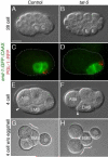
Figure 1. TAT-5 is required for cell shape changes and gastrulation
(A–B) Stills from DIC movie of 28-cell embryos. `E' indicates endodermal precursors. (C–D) Endodermal precursors express end-1GFPCAAX during gastrulation in control (C) and tat-5 (D) 26-cell embryos. P granules (red) localize to the germ cell precursor. (E–F) Stills from DIC movie of late 4-cell embryos. Arrowheads point to the anterior edge of EMS, which extends under ABa in control but not tat-5 embryos. (G–H) Stills from DIC movie of devitellinized late 4-cell embryos. Cells appear more rounded in tat-5 embryos as denoted by contour lines. All images lateral views with anterior to the left and dorsal up. Scale bar is 15 μm. See also Figure S1, Movie S1, and Movie S2.
Figure 2
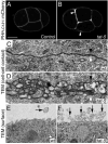
Figure 2. Extracellular vesicles are present on cell surfaces and between cell-cell contacts in tat-5 embryos
(A–B) PHPLC1∂1∷mCherry in 4-cell embryos. Arrowheads point to thick patches of membrane in tat-5 embryos. Scale bar is 15 μm. (C–D) TEM of the cell contact from 2-cell embryos. Black and white arrowheads point to the plasma membrane of different cells. (E–F) TEM of the surface of one-cell embryos. Arrows point to extracellular vesicles or tubules. Scale bars in C–F are 200 nm. See Figure S2 for additional TEMs.
Figure 3
Figure 3. Extracellular vesicles are generated by budding from the plasma membrane
(A) Model based on a TEM tomogram depicting the range in vesicle (arrowhead) and tubulovesicle (arrow) shapes found in a 2-cell tat-5 embryo. Each color represents a distinct structure and only membranes are traced. (A') Box from A depicting a vesicle (shaded) connected to the plasma membrane. See also Movie S3 for vesicle shapes. (B) Histogram of vesicle diameters from tat-5 intralumenal MVB vesicles (light grey) averaging 65 ± 15 nm, n=110 from 3 embryos, and extracellular vesicles (dark grey) averaging 149 ± 35 nm, n=89 ECVs from 3 embryos. (C–D) GFP∷ZF1∷SYX-4 (green) in 26-cell embryos. Dotted lines trace cells where ZF1 degradation has occurred. In insets, SAX-7 (red) labels the plasma membrane. Nuclei are blue. (E–F) Surface view of 26-cell embryos expressing med-1GFPCAAX. Oval outlines embryos. Arrowheads point to membrane thickening. See Figure S3 for exosome marker data.
Figure 4
Figure 4. TAT-5 localizes to the plasma membrane and maintains PE asymmetry
(A–B) GFP∷TAT-5 (A) and GFP∷TAT-5(E246Q) (B) expression in 2-cell embryos. (C–D) GFP∷TAT-5 (C) rescues (n=164 embryos) and GFP∷TAT-5(E246Q) (D) fails to rescue (n=28 embryos) membrane thickenings (arrowhead) in 15-cell embryos from tat-5(tm1741) mothers. (E–G) Annexin V staining in 8–16 cell devitellinized embryos. (H) Graph of normalized Annexin V membrane labeling intensity (Control: n=20 embryos, tat-1: n=14, tat-5: n=14). (I–K) Duramycin staining in 44–46 cell devitellinized embryos. Arrowhead marks a cell contact. (L) Graph of normalized Duramycin membrane labeling intensity (Control: n=15 embryos, tat-1: n=12, tat-5: n=7). Data are represented as mean +/- SD. Compared to control: *p<0.05, **p<0.01. See Figure S4 for GFP∷TAT-5(D439E) localization and overall phospholipid content.
Figure 5
Figure 5. Clathrin, RAB-11, and the ESCRT complex are recruited to the plasma membrane in tat-5 embryos
(A–B) GFP∷CHC-1 (clathrin) in 4-cell embryos. Arrowhead indicates cell contact. (C–D) GFP∷RAB-11 in 4-cell embryos. Bright spot is midbody. (E–F) GFP∷MVB-12 (ESCRT-I) in 4-cell embryos from vps-4 feeding RNAi (fRNAi) (E) and tat-5 + vps-4 dsRNA injections (F). (G–H) Projections of 2-cell embryos stained for ESCRT-III protein VPS-32 (green) from vps-4 fRNAi (G) or tat-5 + vps-4 dsRNA (H) injections (vps-4: 0.27 ± 0.11 puncta/μm, n=7, 136 μm examined; tat-5 + vps-4: 0.62 ± 0.16 puncta/μm, n=4, 97 μm examined; p<0.01). PAR-3 (red) marks cell cortex. See Figure S5 for more endosomal markers.
Figure 6
Figure 6. RAB-11 and the ESCRT complex mediate extracellular vesicle production
(A–B) Graphs displaying percentage of embryos +/- SD with wild-type membranes, or mild or severe thickenings. Thickenings were judged mild if they only affected a subset of three-cell junctions or severe if they affected two- and three-cell junctions. Compared to tat-5: *p<0.05, **p<0.01. (C–H) Embryos expressing PHPLC1∂1∷GFP from adults injected with the indicated dsRNAs. See Figure S6 for controls and Table S1 for n values.
Figure 7
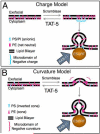
Figure 7. Two models for the role of TAT-5 and PE externalization in ectosome production
(A–B) TAT-5 normally internalizes PE. PE externalization by an unknown scramblase could lead to the recruitment or stabilization of the ESCRT complex to the plasma membrane, where the ESCRT complex buds out ectosomes. RAB-11 could deliver proteins needed for budding to the plasma membrane or could have a more direct role in the budding process. (A) Externalized PE results in microdomains of increased negative charge at the cytoplasmic face (from relatively increased PS and PI density). (B) Externalized PE induces domains of negative membrane curvature that could act as hinge regions. These models are not exclusive.

Comment in

References

    1. Tanaka Y, Okada Y, Hirokawa N. FGF-induced vesicular release of Sonic hedgehog and retinoic acid in leftward nodal flow is critical for left-right determination. Nature. 2005;435:172–177. - PubMed
    1. Gyorgy B, Szabo TG, Pasztoi M, Pal Z, Misjak P, Aradi B, Laszlo V, Pallinger E, Pap E, Kittel A, et al. Membrane vesicles, current state-of-the-art: emerging role of extracellular vesicles. Cell Mol Life Sci. 2011;68:2667–2688. - PMC - PubMed
    1. Théry C, Ostrowski M, Segura E. Membrane vesicles as conveyors of immune responses. Nat Rev Immunol. 2009;9:581–593. - PubMed
    1. Basse F, Gaffet P, Rendu F, Bienvenue A. Translocation of spin-labeled phospholipids through plasma membrane during thrombin- and ionophore A23187-induced platelet activation. Biochemistry. 1993;32:2337–2344. - PubMed
    1. Morris RJ. Ionic control of the metastable inner leaflet of the plasma membrane: Fusions natural and artefactual. FEBS LETTERS. 2009;584:1665–1669. - PubMed

Publication types

MeSH terms