. 2011 Nov 17;10(5):437-50.
doi: 10.1016/j.chom.2011.09.011.

Innate recognition of cell wall β-glucans drives invariant natural killer T cell responses against fungi

Affiliations

Innate recognition of cell wall β-glucans drives invariant natural killer T cell responses against fungi

Nadia R Cohen et al. Cell Host Microbe. .

Abstract

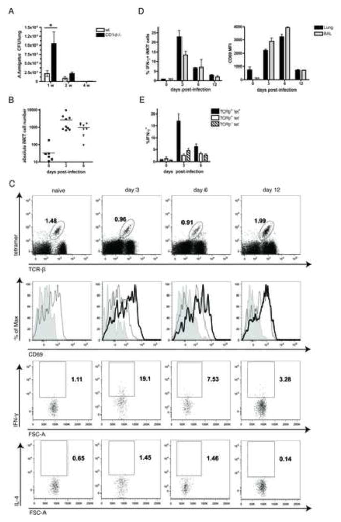
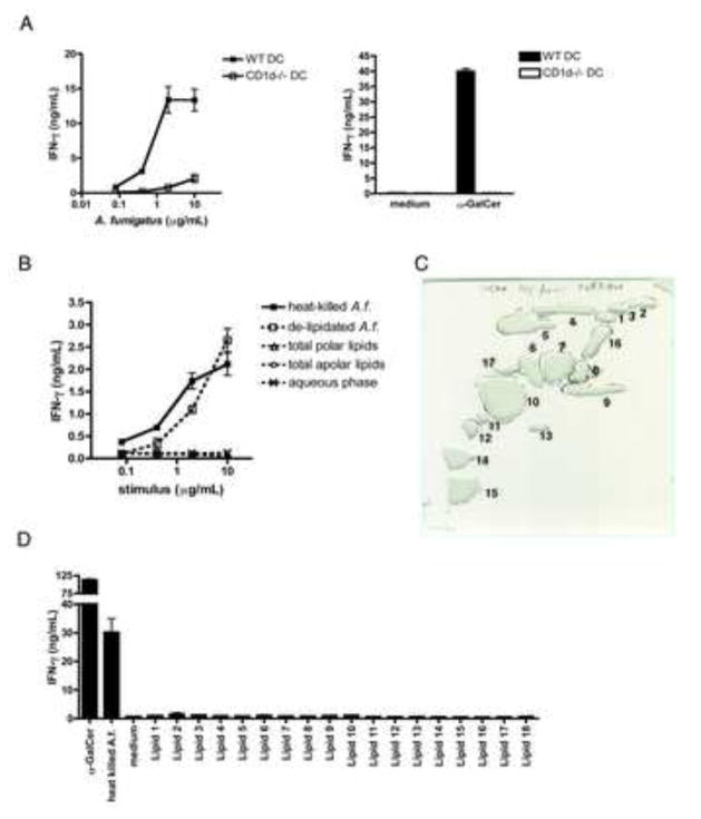
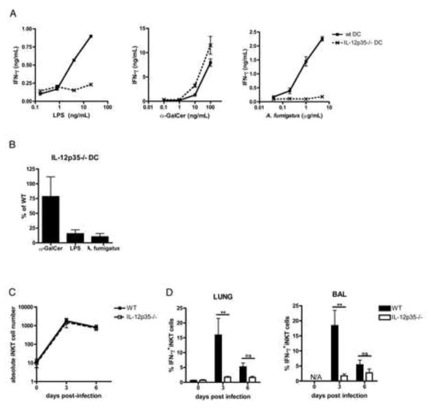
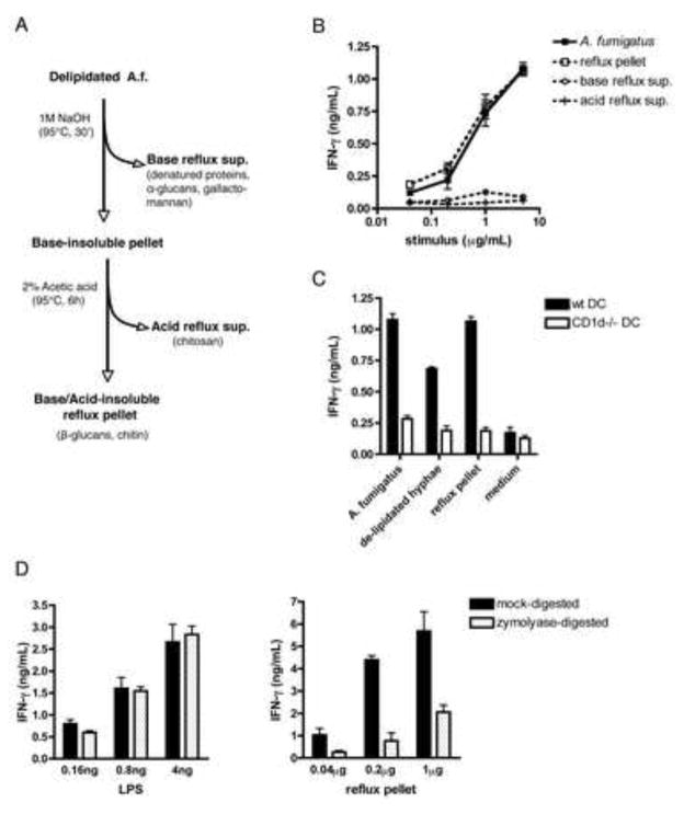
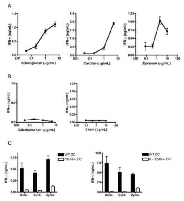
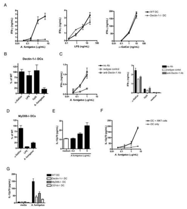
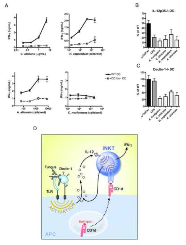
iNKT cells are innate T lymphocytes recognizing endogenous and foreign lipid antigens presented in the MHC-like molecule CD1d. The semi-invariant iNKT cell TCR can detect certain bacterial and parasitic lipids and drive iNKT cell responses. How iNKT cells respond to fungi, however, is unknown. We found that CD1d-deficient mice, which lack iNKT cells, poorly control infection with the fungal pathogen Aspergillus fumigatus. Furthermore, A. fumigatus rapidly activates iNKT cells in vivo and in vitro in the presence of APCs. Surprisingly, despite a requirement for CD1d recognition, the antifungal iNKT cell response does not require fungal lipids. Instead, Dectin-1- and MyD88-mediated responses to β-1,3 glucans, major fungal cell-wall polysaccharides, trigger IL-12 production by APCs that drives self-reactive iNKT cells to secrete IFN-γ. Innate recognition of β-1,3 glucans also drives iNKT cell responses against Candida, Histoplasma, and Alternaria, suggesting that this mechanism may broadly define the basis for antifungal iNKT cell responses.


Conflict of interest statement

The authors have no conflicting financial interests.

Figures

Fig. 1
Fig. 1. CD1d-restricted T cells participate in the immune response against A.f
WT or CD1d−/− B6 mice were infected i.t. with 1×10⁷ (a) or 4×10⁷ (b–e) live A.f. conidia and euthanized at the indicated timepoints. (a) Fungal CFU in lung homogenates. Bars, mean ± SEM; n=4–5 mice/group, data representative of 3 independent experiments. (b) Absolute number of iNKT cells (CD45+, CD19-, TCR-β+, α-GalCer-loaded CD1d tetramer+) in the BAL. Data pooled from 3 independent experiments. (c) Lung iNKT cell percentage (top row), CD69 MFI (second row) and cytokine secretion (bottom rows) following infection. Shaded histogram, isotype control; grey line, naïve mouse; black line, infected mouse. (d) Average %IFN-γ+ iNKT and CD69 MFI following infection. N/A, not applicable (to few cells). Bars, mean ± SEM, n=3 mice. Data representative of ≥ 4 independent experiments. (e) Average %IFN-γ+ iNKT cells, tet T cells, and NK cells, (TCR-βtet) following infection. Data pooled from 4 independent experiments, Bars, mean ± SEM, n=9–12 mice.
Fig. 2
Fig. 2. Non-lipidic components of the A.f. cell wall drive iNKT cell activation in vitro
105 iNKT cells and 104 CD11c+ DCs were co-cultured for 16–20hrs with the indicated stimuli. IFN-γ in the supernatants was measured by ELISA. (a) IFN-γ secreted in response to heat-killed A.f. hyphae, α-GalCer (100ng/mL) or medium in co-cultures of iNKT cells and WT or CD1d−/− DCs. (b) IFN-γ secreted in response to lipidic and non-lipidic fungal fractions extracted from hyphae in co-cultures of iNKT cells and WT DCs. (c) Preparative 2D-TLC purification of A.f. polar lipids. (d) IFN-γ secreted in response to α-GalCer (10ng/mL), A.f. (20μg/mL) or 2D TLC-purified polar lipid species (20μg/mL) in co-cultures containing iNKT cells and WT DCs. (a–d) Error bars, ± SEM, data representative of ≥ 3 independent experiments.
Fig. 3
Fig. 3. A.f.-driven iNKT cell activation is IL-12p70-dependent in vitro and in vivo
(a) IFN-γ in the supernatant of co-cultures containing iNKT cells and WT or IL-12p35−/− DCs in the presence of the indicated stimuli, error bars, ± SEM. (b) Average IFN-γ in supernatants of co-cultures containing α-GalCer (100ng/mL), LPS (20ng/mL) or A.f. (5μg/mL), iNKT cells and IL-12p35−/− DCs as a percentage of the IFN-γ in co-cultures containing WT DCs, data pooled from 3 independent experiments, error bars, ± SEM. (c) Absolute number and (d) average %IFN-γ-secreting iNKT cells in the airways of WT or IL-12p35−/− mice post-infection. Bars, mean ± SEM, n=5–6 mice, data pooled from 2 independent experiments.
Fig. 4
Fig. 4. Cell wall β1,3-glucans are responsible for A.f.-driven iNKT cell activation in vitro
(a) Fungal polysaccharide fractionation protocol. (b) Activity of reflux supernatants and pellet in co-culture assay containing iNKT cells and WT DCs. (c) IFN-γ secretion in co-cultures containing iNKT cells, either wild type or CD1d−/− DC and 5μg/mL of the indicated fractions. (d) IFN-γ secretion in co-cultures containing LPS or the A.f. reflux pellet digested overnight with zymolyase. (a–d) Error bars, ± SEM, data representative of 2–4 independent experiments.
Fig. 5
Fig. 5. Microbial β-glucans activate iNKT cells in the presence of CD1d- and IL-12-sufficient APCs
(a–c) IFN-γ secretion in iNKT cells co-cultures containing the indicated polysaccharides in increasing concentrations (a, b) or at 2–5μg/mL (c), error bars, ± SEM, data representative of 2–4 independent experiments.
Fig. 6
Fig. 6. A.f.-driven iNKT cell activation depends on APC-mediated fungal recognition via the C-type lectin Dectin-1 and MyD88 signaling
(a) IFN-γ detected in the supernatants of co-cultures containing iNKT cells, WT or Dectin-1−/− DCs and the indicated stimuli. (b) Average IFN-γ in iNKT cell co-cultures containing α-GalCer (10ng/mL), CpG (1μg/mL), LPS (4ng/mL) or A.f. (5μg/mL) and Dectin-1−/− DCs or (d) MyD88−/− DCs as a percentage of the IFN-γ measured in co-cultures containing WT DCs, data pooled from 3 independent experiments. (c) Co-culture performed in the presence of 2.5μg/mL of anti-Dectin-1 blocking mAb, an isotype control, or no antibody, and heat-killed A.f. hyphae (left), α-GalCer (10ng/mL), CpG (1μg/mL) or medium (right). Data representative of 2 independent experiments. (e) IL12p70 detected in the supernatants of WT DCs co-cultured with the indicated amounts of heat-killed A.f. in the absence or (f) presence of iNKT cells. (g) IL12p70 detected in the supernatants of co-cultures containing DCs, iNKT cells and either medium or A.f. (5μg/mL). Data representative of 2–3 independent experiments. (a–g) Error bars, ± SEM.
Fig. 7
Fig. 7. Dectin-1-mediated recognition of pathogenic fungi and IL-12 secretion by activated APCs drives iNKT cell activation
(a) IFN-γ detected in the supernatants of co-cultures containing iNKT cells, WT or CD1d−/− DCs and heat-killed fungi. (b) Average IFN-γ in iNKT cell co-cultures containing α-GalCer (1ng/mL), LPS (20ng/mL), A.f. (5μg/mL), C.a. (5μg/mL), H.c. (8–40×103 cells/well), A.a. (1.6×10³ cells/well) and IL-12p35−/− or (c) Dectin-1−/− DCs as a percentage of the IFN-γ in co-cultures containing WT DCs, data pooled from 3–4 independent experiments. (a–c) Error bars, ± SEM. (d) Model mechanism for fungal-driven iNKT cell activation.

Comment in

  • iNKTs foil fungi.
    Prlic M, Hohl TM. Prlic M, et al. Cell Host Microbe. 2011 Nov 17;10(5):421-2. doi: 10.1016/j.chom.2011.11.003. Cell Host Microbe. 2011. PMID: 22100157 Free PMC article.

References

    1. Amprey JL, Im JS, Turco SJ, Murray HW, Illarionov PA, Besra GS, Porcelli SA, Spath GF. A subset of liver NK T cells is activated during Leishmania donovani infection by CD1d-bound lipophosphoglycan. J Exp Med. 2004;200:895–904. - PMC - PubMed
    1. Balloy V, Chignard M. The innate immune response to Aspergillus fumigatus. Microbes Infect. 2009;11:919–927. - PubMed
    1. Bendelac A, Savage PB, Teyton L. The biology of NKT cells. Annu Rev Immunol. 2007;25:297–336. - PubMed
    1. Bernard M, Latge JP. Aspergillus fumigatus cell wall: composition and biosynthesis. Med Mycol. 2001;39(Suppl 1):9–17. - PubMed
    1. Brakhage AA, Bruns S, Thywissen A, Zipfel PF, Behnsen J. Interaction of phagocytes with filamentous fungi. Curr Opin Microbiol. 2010;13:409–415. - PubMed

Publication types