. 2011 Dec 13;21(6):1014-25.
doi: 10.1016/j.devcel.2011.09.010. Epub 2011 Nov 17.

Conditional deletion of Msx homeobox genes in the uterus inhibits blastocyst implantation by altering uterine receptivity

Affiliations

Conditional deletion of Msx homeobox genes in the uterus inhibits blastocyst implantation by altering uterine receptivity

Takiko Daikoku et al. Dev Cell. .

Abstract

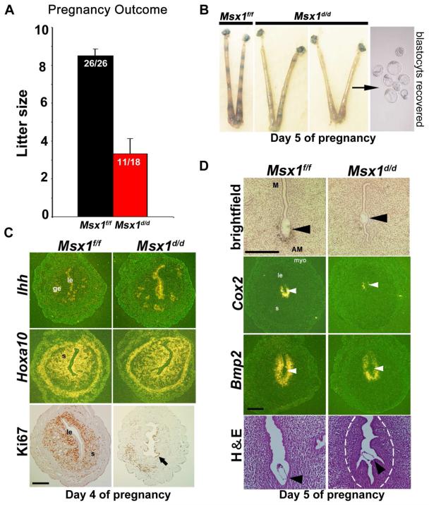
An effective bidirectional communication between an implantation-competent blastocyst and the receptive uterus is a prerequisite for mammalian reproduction. The blastocyst will implant only when this molecular cross-talk is established. Here we show that the muscle segment homeobox gene (Msh) family members Msx1 and Msx2, which are two highly conserved genes critical for epithelial-mesenchymal interactions during development, also play crucial roles in embryo implantation. Loss of Msx1/Msx2 expression correlates with altered uterine luminal epithelial cell polarity and affects E-cadherin/β-catenin complex formation through the control of Wnt5a expression. Application of Wnt5a in vitro compromised blastocyst invasion and trophoblast outgrowth on cultured uterine epithelial cells. The finding that Msx1/Msx2 genes are critical for conferring uterine receptivity and readiness to implantation could have clinical significance, because compromised uterine receptivity is a major cause of pregnancy failure in IVF programs.


Figures

Figure 1
Figure 1. Msx1 is expressed in a spatiotemporal manner in the uterus and is influenced by LIF
(A) β-gal staining of Msx1-LacZ in uterine sections on days 1-5 of pregnancy and on day 6 of pseudopregnancy (pDay 6). Sections were counterstained with eosin. Arrowhead denotes the implanting blastocyst. Bar, 500μm. (B) Msx1 expression in ovariectomized, delayed implanting uteri before and after E2 activation. Arrowheads denote the location of unattached (delayed) or attached (activated) blastocysts in longitudinal uterine sections. Bar, 400μm. (C) Northern hybridization of Msx1 in ovariectomized WT and Lif−/− mice treated with P4, P4 + E2, or P4 + recombinant LIF (rLIF). (D&E) Northern hybridization of Msx1 and Lif in ovariectomized pseudopregnant WT uteri treated with P4+E2 or P4, and quantification of their relative expression levels. (F) In situ hybridization of Msx1 in day 6 pseudopregnant uteri conditionally deleted of Gp130. Bar, 250μm. le, luminal epithelium; ge, glandular epithelium; s, stroma; myo, myometrium.
Figure 2
Figure 2. Msx1d/d mice show compromised pregnancy due to failed or defective implantation
(A) Pregnancy success and litter sizes in Msx1f/f and Msx1d/d females (mean ± SEM). (B) Defective or failed implantation in Msx1d/d mice as determined by blue dye injection. Blastocysts were recovered from uteri with no or weak blue bands. (C) In situ hybridization of Ihh and Hoxa10, and immunostaining of Ki67 on day 4 of pregnancy. Arrow denotes epithelial staining. Bar, 400 μm. (D) In situ hybridization of Cox2 and Bmp2 and uterine histology on day 5. Arrowheads denote location of implanting blastocysts. M, mesometrial end; AM, anti-mesometrial end. Demarcated area within the H&E stained section depicts reduced edema. Bars, 400 μm (in situ hybridization) and 100 μm (brightfield and H&E stained sections). See also Figure S1, and Tables S1A and S2A
Figure 3
Figure 3. Defective implantation in Msx1d/d females with poor embryo spacing and decidualization with increased resorption rates
(A-C) Representative uteri from Msx1f/f and Msx1d/d females on days 6, 8 and 12. Arrowheads denote resorption sites, whereas an arrow shows normal implantation. (D) Resorption rate on day 12 in Msx1d/d females. Numbers within parentheses indicate number of resorptions over total number of implantation sites. Number of females examined are shown on top of the bars. (E) Decidualization in Msx1d/d mice after intrauterine oil infusion. The right horn received oil infusion, while the left horn served as a control. (F) Fold changes in uterine weights of oil-infused horns over non-infused horns indicate the extent of decidualization (mean ± SEM). See also Figure S2.
Figure 4
Figure 4. Deletion of both Msx1 and Msx2 leads to complete infertility with aberrant expression of implantation-specific genes
(A) Northern hybridization of uterine Msx2 on days 1-4 in WT females. (B) In situ hybridization of Msx2 in Msx1d/d mice. Bar, 400 μm. (C) RT-PCR of Msx2 in Msx1d/d uteri. (D) Msx1/Msx2d/d females are totally infertile (mean ± SEM). (E) Implantation failure in Msx1/Msx2d/d females on day 5 (mean ± SEM). (F) In situ hybridization of Bmp2 and Cox2 in uterine sections at the site of blastocyst apposition. Arrowheads denote locations of blastocysts. Bars, 400 μm (darkfield) and 100 μm (brightfield), respectively. See also Figure S3, and Tables S1B and S2B
Figure 5
Figure 5. Uterine Lif expression is downregulated in mice deleted of uterine Msx genes and LIF administration fails to rescue implantation
(A) Lif expression in Msx1f/f and Msx1d/d uteri. Arrowheads denote the locations of blastocysts. Bar, 400 μm. (B) Lif expression in Msx1/Msx2f/f and Msx1/Msx2d/d uteri. Bar, 400μm. (C) Implantation of transferred blastocysts in Lif−/−/Msx1f/f and Lif−/−/Msx1d/d recipients after injection of LIF. Implantation sites (IS) were examined on days 5 or 6. Unimplanted blastocysts recovered from uteri confirmed successful transfer. Parentheses indicate the ratio of implantation sites to transferred blastocysts (mean ± SEM). (D) Representative images Lif−/−/Msx1f/f or Lif−/−/Msx1d/d uteri after blue dye injection and recovered blastocysts. (E) Implantation in Lif−/− and Msx1/Msx2d/d females (n=3) after LIF injection on day 5. Uteri without IS were flushed to recover blastocysts to confirm pregnancy. See also Tables S3A & B.
Figure 6
Figure 6. Msx genes regulate epithelial cell polarity via Wnt5a
(A) Western blotting of uterine β-catenin, E-cadherin and Wnt5a. (B) Confocal images of E-cadherin and β-catenin co-localization. Retention of the LE in Msx1/Msx2d/d mice at the site of blastocyst apposition on day 6, as opposed to LE breakdown in Msx1/Msx2f/f mice with loss of E-cadherin. Arrowhead indicates the location of blastocysts. Bar, 100μm. (C) Confocal images of E-cadherin and β-catenin co-localization at the inter-implantation sites in Msx1/Msx2f/f and Msx1/Msx2d/d females on day 6. Blue, nuclear stain. Bar, 100μm. (D) Co-IP of E-cadherin/β-catenin in primary endometrial cells of Msx1/Msx2f/f and Msx1/Msx2d/d mice on day 5. Ratio of immunoprecipitated E-cadherin to β-catenin was determined for Msx1/Msx2f/f and Msx1/Msx2d/d endometria and normalized to that of Msx1/Msx2f/f endometrium (right panel). (E&F) Immunohistochemistry of acetylated α-tubulin (E) and ezrin (F) in Msx1/Msx2f/f and Msx1/Msx2d/d uteri. bv, blood vessels. Bars, 100μm. (G) ChIP showing enriched binding of Msx1 to the Wnt5a promoter region. See also Figure S4.
Figure 7
Figure 7. Wnt5a compromises blastocyst outgrowth on uterine epithelial cells
(A) Confocal images of E-cadherin and β-catenin co-localization in cultured primary epithelial cells in the presence or absence of Wnt5a. Bar, 100μm. (B&C) Blastocysts cultured on primary epithelial cells (LE) atop stromal cells (B) or separated from stromal cells (S) by an insert (C) in the presence or absence of Wnt5a. (D) Proposed model of regulation of implantation by Msx genes. Higher levels of Wnt5a in the absence of Msx1 and/or Msx2 confer higher luminal epithelial polarity by enhancing E-cadherin/β-catenin complex formation, impeding the breakdown of the epithelial barrier for implantation. See also Figure S5.

References

    1. Bach A, Lallemand Y, Nicola MA, Ramos C, Mathis L, Maufras M, Robert B. Msx1 is required for dorsal diencephalon patterning. Development. 2003;130:4025–4036. - PubMed
    1. Bei M, Maas R. FGFs and BMP4 induce both Msx1-independent and Msx1-dependent signaling pathways in early tooth development. Development. 1998;125:4325–4333. - PubMed
    1. Betz UA, Bloch W, van den Broek M, Yoshida K, Taga T, Kishimoto T, Addicks K, Rajewsky K, Muller W. Postnatally induced inactivation of gp130 in mice results in neurological, cardiac, hematopoietic, immunological, hepatic, and pulmonary defects. J Exp Med. 1998;188:1955–1965. - PMC - PubMed
    1. Catron KM, Wang H, Hu G, Shen MM, Abate-Shen C. Comparison of MSX-1 and MSX-2 suggests a molecular basis for functional redundancy. Mech Dev. 1996;55:185–199. - PubMed
    1. Cha SW, Tadjuidje E, Tao Q, Wylie C, Heasman J. Wnt5a and Wnt11 interact in a maternal Dkk1-regulated fashion to activate both canonical and non-canonical signaling in Xenopus axis formation. Development. 2008;135:3719–3729. - PubMed

Publication types

MeSH terms