. 2011 Dec 16;416(3-4):293-9.
doi: 10.1016/j.bbrc.2011.11.028. Epub 2011 Nov 12.

Increased cardiogenesis in P19-GFP teratocarcinoma cells expressing the propeptide IGF-1Ea

Affiliations

Increased cardiogenesis in P19-GFP teratocarcinoma cells expressing the propeptide IGF-1Ea

Bhawana Poudel et al. Biochem Biophys Res Commun. .

Abstract

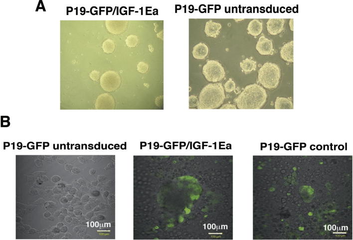
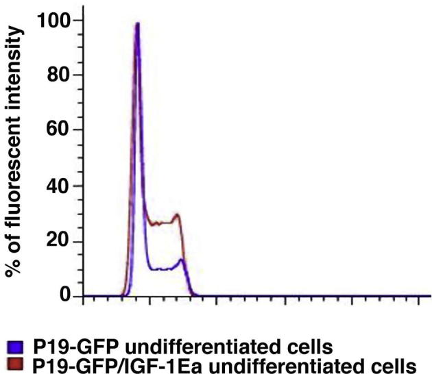
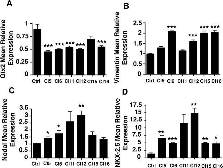
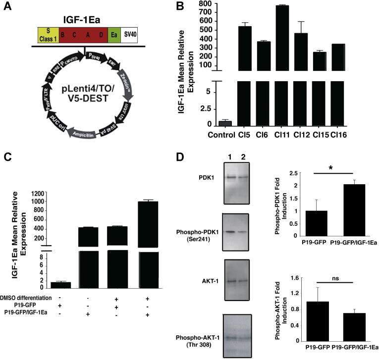
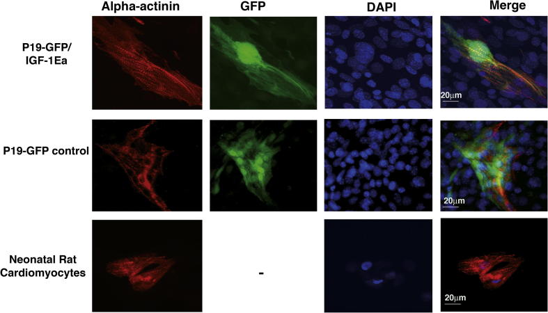
The mechanism implicated in differentiation of endogenous cardiac stem cells into cardiomyocytes to regenerate the heart tissue upon an insult remains elusive, limiting the therapeutical goals to exogenous cell injection and/or gene therapy. We have shown previously that cardiac specific overexpression of the insulin-like growth factor 1 propeptide IGF-1Ea induces beneficial myocardial repair after infarct. Although the mechanism is still under investigation, the possibility that this propeptide may be involved in promoting stem cell differentiation into the cardiac lineage has yet to be explored. To investigate whether IGF-1Ea promote cardiogenesis, we initially modified P19 embryonal carcinoma cells to express IGF-1Ea. Taking advantage of their cardiomyogenic nature, we analyzed whether overexpression of this propeptide affected cardiac differentiation program. The data herein presented showed for the first time that constitutively overexpressed IGF-1Ea increased cardiogenic differentiation program in both undifferentiated and DMSO-differentiated cells. In details, IGF-1Ea overexpression promoted localization of alpha-actinin in finely organized sarcomeric structure compared to control cells and upregulated the cardiac mesodermal marker NKX-2.5 and the ventricular structural protein MLC2v. Furthermore, activated IGF-1 signaling promoted cardiac mesodermal induction in undifferentiated cells independently of cell proliferation. This analysis suggests that IGF-1Ea may be a good candidate to improve both in vitro production of cardiomyocytes from pluripotent stem cells and in vivo activation of the differentiation program of cardiac progenitor cells.


Figures

Supplementary Figure 1
Supplementary Figure 2
Supplementary Figure 3
Fig. 1
Fig. 1
Characterization of P19-GFP embryonal carcinoma cells expressing IGF-1Ea. (A) The pLenti4/TO/V5-DEST contains the recombination sites attR1 and attR2 downstream the CMV-TO promoter that allowed recombination between this lentiviral vector and the one carrying IGF1Ea. (B) Transduced clones constitutively expressed IGF-1Ea transcripts. IGF-1 expression was analyzed by a rodent IGF-1 probe. 18S rRNA was used as internal reference gene. Values are Mean +/− SEM. (C) IGF-1Ea Clone 6 cells and control Clone 6 cells were differentiated in 1% DMSO and RNA extracts analyzed for quantitative Real Time PCR (qRT-PCR). 18S rRNA was used as internal reference gene. Results are expressed as mean ± SEM. (D) Undifferentiated P19-GFP cells expressing IGF-1Ea (lane 1) and control cells (lane 2) were analyzed for PDK1 and AKT1 activity. Normalization was performed with total AKT1 and PDK1. The figure shows the analysis of three independent experiments and their fold induction values in the right panel. Asterisk () indicates significant values (p < 0.05); ns, no significant.
Fig. 2
Fig. 2
IGF-1Ea regulation of survival, proliferation and lineage commitment in undifferentiated P19-GFP cells. (A) P19-GFP/IGF-1Ea and control P19-GFP cells were exposed to 3% hypoxia for 24 h. Percentage of LDH activity was measured as described in Supplementary Materials and Methods by comparison of IGF-1Ea and control cells in normoxic (white bars) and hypoxic (black bars) conditions. Significance was measured by one-way ANOVA and ∗∗ indicates p < 0.01, whereas ∗∗∗ indicates p < 0.001. (B) Cell cycle analysis of undifferentiated P19Cl6 cells, transduced with empty (blue) or IGF-1Ea (red) viral vectors. Percentage of cells in S phase were similar in both empty and IGF-1Ea expressing cells. Results were obtained by PI staining using Flow Jo analyses. X-axis represents percentage of cells and y-axis fluorescent intensity. (C–G) qRT-PCR analysis for the mesodermal markers vimentin (C) and nodal (F), the ectodermal marker Otx2 (D), the endodermal GATA4 (E) and the cardiac marker NKX-2.5 (F) in undifferentiated IGF-1Ea and control cells. 18S rRNA was used as internal reference gene. Results are expressed as mean ± SEM. Asterisk () indicates significant values (p < 0.05); ∗∗ indicates p < 0.01, whereas ∗∗∗ indicates p < 0.001. (For interpretation of the references in color in this figure legend, the reader is referred to the web version of this article.)
Fig. 3
Fig. 3
Differentiation program mediated by IGF-1Ea. (A) Percentage of GFP positive cells expressing IGF-1Ea and control cells quantified by FACS analysis. The analysis was performed in five independent experiments. Values are mean ± SEM. Significance was analyzed by Student’s t-test and p = 0.52. (B–D) qRT-PCR analysis for cardiac specific marker expression in IGF-1Ea and control cells in DMSO-differentiation conditions. (B) Nkx2.5, (C) MLC2v and (D) GATA4 expression were normalized by measuring levels of 18S rRNA.
Fig. 4
Fig. 4
IGF-1Ea mediated cardiomyocytes sarcomeric structure maturation. IGF-1Ea transduced cells (P19-GFP/IGF-1Ea), control cells transduced with lentivirus not expressing IGF-1Ea (P19-GFP) were cultured after embryoid bodies’ formation on 0.1% gelatin. Cardiomyocytes (GFP labeled panels) were stained with alpha actinin monoclonal antibody (Sigma). Nuclei were visualized by DAPI staining. Neonatal rat cardiomyocytes were used as positive control (lower panels). The measurement bar is 20 μm. The pictures are representative of two independent experiments.

Similar articles

Cited by

References

    1. Sutton M.G., Sharpe N. Left ventricular remodeling after myocardial infarction: pathophysiology and therapy. Circulation. 2000;101:2981–2988. - PubMed
    1. Capi O., Gepstein L. Myocardial regeneration strategies using human embryonic stem cell-derived cardiomyocytes. J. Control Release. 2006;116:211–218. - PubMed
    1. Naito A.T., Shiojima I., Akazawa H., Hidaka K., Morisaki T., Kikuchi A., Komuro I. Developmental stage-specific biphasic roles of Wnt/beta-catenin signaling in cardiomyogenesis and hematopoiesis. Proc. Natl. Acad. Sci. USA. 2006;103:19812–19817. - PMC - PubMed
    1. Ueno S., Weidinger G., Osugi T., Kohn A.D., Golob J.L., Pabon L., Reinecke H., Moon R.T., Murry C.E. Biphasic role for Wnt/beta-catenin signaling in cardiac specification in zebrafish and embryonic stem cells. Proc. Natl. Acad. Sci. USA. 2007;104:9685–9690. - PMC - PubMed
    1. Takahashi T., Lord B., Schulze P.C., Fryer R.M., Sarang S.S., Gullans S.R., Lee R.T. Ascorbic acid enhances differentiation of embryonic stem cells into cardiac myocytes. Circulation. 2003;107:1912–1916. - PubMed

Publication types

Substances

LinkOut - more resources