Review
. 2012 Jan;17(1):1-8.
doi: 10.1016/j.tplants.2011.10.005. Epub 2011 Nov 17.

Chloroviruses: not your everyday plant virus

Affiliations
Review

Chloroviruses: not your everyday plant virus

James L Van Etten et al. Trends Plant Sci. 2012 Jan.

Abstract

Viruses infecting higher plants are among the smallest viruses known and typically have four to ten protein-encoding genes. By contrast, many viruses that infect algae (classified in the virus family Phycodnaviridae) are among the largest viruses found to date and have up to 600 protein-encoding genes. This brief review focuses on one group of plaque-forming phycodnaviruses that infect unicellular chlorella-like green algae. The prototype chlorovirus PBCV-1 has more than 400 protein-encoding genes and 11 tRNA genes. About 40% of the PBCV-1 encoded proteins resemble proteins of known function including many that are completely unexpected for a virus. In many respects, chlorovirus infection resembles bacterial infection by tailed bacteriophages.


Figures

Figure 1
Figure 1
Chlorella cells and chlorovirus PBCV-1. (a) Plaques formed by PBCV-1 on a lawn of C. variabilis. (b) Paramecium bursaria and its symbiotic chlorella cells (size bar: 20 μm). (c) C. variabilis cells cultured in the laboratory free of both Paramecium and virus (size bar: 50 μm). (d) Thin section of a healthy C. variabilis cell (size bar: 1 μm). (e) Five-fold averaged cryo-electron micrograph of PBCV-1 reveals a long narrow cylindrical spike structure at one vertex (orange) and fibers extending from one unique capsomer per trisymmetron. The virion has a diameter varying from 1,650 Å, measured along the 2-fold and 3-fold axes, and 1,900 Å, measured along the 5-fold axes. The glycoprotein shell is composed of 20 triangular units, or “trisymmetrons,” and 12 pentagonal caps, or “pentasymmetrons,” at the 5-fold vertices (yellow). (f) Central cross section of (e). Note the gap between the unique vertex and the membrane enclosing the DNA. (g) Surface view of the PBCV-1 spike structure and fibers. (h) A magnified image of the spike structure (in blue). Also note a plug structure (in pink) inside the spike (size bar: 20 nm). (i) PBCV-1 attached to the cell wall as viewed by the quick-freeze, deep etch procedure. Note the virions attached to the wall by fibers. (j) Initial attachment of PBCV-1 to C. variabilis cell walls. The orientation of the unique vertex (recognized by the pocket under the vertex) can be seen for the two virus particles marked with red arrows. These virus particles have their unique vertex facing the cell wall indicating that these particles recognize the cell surface. (k) Attachment of PBCV-1 to the algal wall and digestion of the wall at the point of attachment. This occurs within 1-3 min p.i. (size bar: 100 nm) (l) Virion particles assemble in defined areas in the cytoplasm named virus assembly centers. Note both DNA containing (dark centers) and empty capsids (size bar: 500 nm). (m) Localized lysis of cell plasma membrane and cell wall and release of progeny viruses at ~8 hr p.i. (size bar: 1 μm). (n/o) C. variabilis cell wall surface of uninfected (n) and 4 hr after PBCV-1 infection (o), as viewed by quick-freeze, deep-etch electron microscopy. Note the accumulation of a dense, fibrous hyaluronan network on the surface of the infected cells. Figure b was kindly provided by Wim van Egmond; Figure e from [13]; Figure f was kindly provided by Xinzheng Zhang and Michael Rossmann; Figures g, h and j from [14]; Figure i from [26]; Figure k from [83], Figures l and m from [84], and Figures n and o from [56] - all published with permission.
Figure 2
Figure 2
Proposed replication cycle of PBCV-1. The virus uncoats at the surface of the alga and the viral DNA, possibly with associated proteins, is assumed to move to the nucleus where early gene transcription begins with 5 to 10 min p.i. The early mRNAs are transported to the cytoplasm for translation, and at least some early proteins presumably return to the nucleus to initiate viral DNA replication, which begins 60 to 90 min p.i., followed by late gene transcription. Late mRNAs are transported to the cytoplasm for translation and many of these late proteins are targeted to the virus assembly centers, where virus capsids are formed and DNA is packaged. The chlorella cell membrane and wall lyses, and infectious PBCV-1 progeny viruses are released at 6 to 8 hr p.i. ( →) Known events; (---->) hypothesized events.
Figure 3
Figure 3
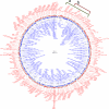
Map of the re-sequenced and annotated PBCV-1 genome (GenBank accession number JF411744.1) presented as a circle. However the genome is a linear molecule and the ends are depicted at the top (12 o'clock) of the figure as a crooked black line. Sequences to the right of the black line are numbered from 1 to 330611 base pairs. Open reading frames are indicated by the block arrows. Those pointing clockwise (red) are annotated with a “R”, those pointing counter-clockwise (blue) are annotated with a “L”. The gene tags are annotated with the prefix “a” indicating the ORF is unlikely to be expressed, or with “A” indicating that it is likely to be expressed, and in most cases shown to be expressed at the RNA level and in many cases as proteins. The polycistronic tRNA gene is located near “6 o'clock”. The two brackets on the top-right of the figure depict two classes of deletion mutants; (a) deletion of map position 4.9 to 42.2, (b) deletion of map position 16 to 42.2. The figure was composed using CGView [85]

Similar articles

Cited by

References

    1. Hull R. Comparative Plant Virology. Elsevier/Academic Press; Amsterdam: 2009.
    1. Nagasaki K. Dinoflagellates, diatoms, and their viruses. J. Microbiol. 2008;46:235–243. - PubMed
    1. Fuhrman JA. Marine viruses and their biogeochemical and ecological effects. Nature. 1999;399:541–548. - PubMed
    1. Suttle CA. Marine viruses - major players in the global ecosystem. Nat. Rev. Microbiol. 2005;5:801–812. - PubMed
    1. Wommack KE, Colwell RR. Viroplankton: viruses in aquatic ecosystems. Microbiol. Mol. Biol. Rev. 2000;64:69–114. - PMC - PubMed

Publication types

MeSH terms

LinkOut - more resources