Review
. 2012 Jun;1818(6):1526-35.
doi: 10.1016/j.bbamem.2011.11.004. Epub 2011 Nov 9.

VDAC inhibition by tubulin and its physiological implications

Affiliations
Review

VDAC inhibition by tubulin and its physiological implications

Tatiana K Rostovtseva et al. Biochim Biophys Acta. 2012 Jun.

Abstract

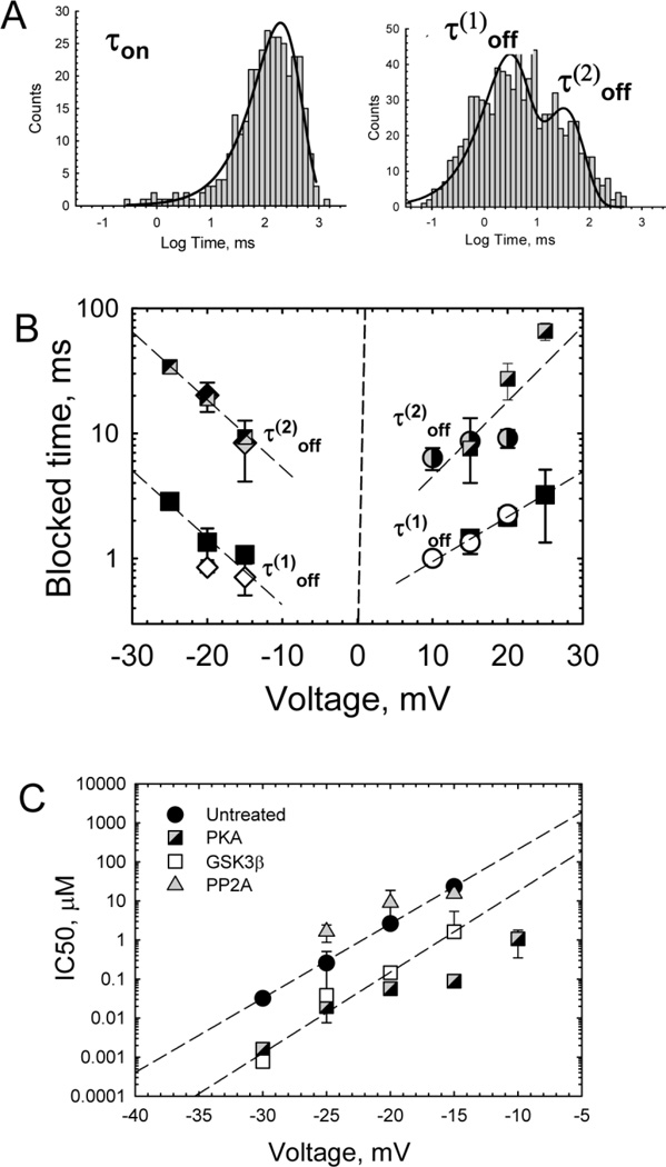
Regulation of mitochondrial outer membrane (MOM) permeability has dual importance: in normal metabolite and energy exchange between mitochondria and cytoplasm, and thus in control of respiration, and in apoptosis by release of apoptogenic factors into the cytosol. However, the mechanism of this regulation involving the voltage-dependent anion channel (VDAC), the major channel of MOM, remains controversial. For example, one of the long-standing puzzles was that in permeabilized cells, adenine nucleotide translocase is less accessible to cytosolic ADP than in isolated mitochondria. Still another puzzle was that, according to channel-reconstitution experiments, voltage regulation of VDAC is limited to potentials exceeding 30mV, which are believed to be much too high for MOM. We have solved these puzzles and uncovered multiple new functional links by identifying a missing player in the regulation of VDAC and, hence, MOM permeability - the cytoskeletal protein tubulin. We have shown that, depending on VDAC phosphorylation state and applied voltage, nanomolar to micromolar concentrations of dimeric tubulin induce functionally important reversible blockage of VDAC reconstituted into planar phospholipid membranes. The voltage sensitivity of the blockage equilibrium is truly remarkable. It is described by an effective "gating charge" of more than ten elementary charges, thus making the blockage reaction as responsive to the applied voltage as the most voltage-sensitive channels of electrophysiology are. Analysis of the tubulin-blocked state demonstrated that although this state is still able to conduct small ions, it is impermeable to ATP and other multi-charged anions because of the reduced aperture and inversed selectivity. The findings, obtained in a channel reconstitution assay, were supported by experiments with isolated mitochondria and human hepatoma cells. Taken together, these results suggest a previously unknown mechanism of regulation of mitochondrial energetics, governed by VDAC interaction with tubulin at the mitochondria-cytosol interface. Immediate physiological implications include new insights into serine/threonine kinase signaling pathways, Ca(2+) homeostasis, and cytoskeleton/microtubule activity in health and disease, especially in the case of the highly dynamic microtubule network which is characteristic of cancerogenesis and cell proliferation. In the present review, we speculate how these findings may help to identify new mechanisms of mitochondria-associated action of chemotherapeutic microtubule-targeting drugs, and also to understand why and how cancer cells preferentially use inefficient glycolysis rather than oxidative phosphorylation (Warburg effect). This article is part of a Special Issue entitled: VDAC structure, function, and regulation of mitochondrial metabolism.


Figures

Figure 1
Figure 1
Tubulin induces fast reversible events of partial blockage of VDAC, which differ from voltage-induced gating of the channel by kinetic parameters and conductance distribution. (A), A representative trace of ion current through a single channel before (left trace) and after (right trace) addition of 50 nM tubulin at −25 mV applied voltage. Tubulin induces a well-defined blocked state. Time-resolved blockage events are shown in Inset at a finer scale. (B), Typical voltage gating of VDAC in the absence of tubulin at −50 mV applied voltage. Voltage of this magnitude moves the channel from a single high-conducting open state to the variety of the low-conducting “closed” states. VDAC was isolated from N. crassa mitochondria. Bilayer membranes were formed from diphytanoyl phosphatidylcholine. Membrane bathing solution contained 1 M KCl with 5 mM HEPES at pH 7.4. The dashed lines indicate zero current. For the sake of presentation convenience, ion current was defined as positive when cations moved from the trans to cis side of the membrane. The records were filtered using an averaging time of 10 ms (except for 1 ms in Inset). (From [10]).
Figure 2
Figure 2
Kinetic analysis of blockage events. (A), The distribution of times between blockage events, when the channel stays open (τon), is satisfactorily described by single exponential fitting. Distributions of the times spent by the channel in the blocked state require at least two exponents for fitting with characteristic times τ(1)off and τ(2)off . (B), Both characteristic times in the blocked state depend symmetrically on the applied voltage when tubulin is added to both sides (squares) or to either side of the membrane when the applied potential is more negative from the side of tubulin addition (cis side addition, diamonds; trans side addition, circles). (C), Voltage dependence of tubulin inhibitory concentration, IC50 (from [54]). IC50 strongly depends on the applied voltage and VDAC phosphorylation state. VDAC was isolated from mouse liver mitochondria with Triton x-100 and then phosphorylated by PKA or GSK3β, or dephosphorylated by PP2A. The IC50 values are averages over datasets obtained in 5–8 experiments with different VDAC samples. The dashed lines are fits to IC50 (V) = IC50(0) exp(nVF/RT), where V is the applied voltage, with the “effective gating charge” n = 11.2 and 12.2 for untreated and GSK3β phosphorylated VDAC, respectively. Other experimental conditions were as in Fig. 1.
Figure 3
Figure 3
A tentative model of the restricted permeation block of VDAC by tubulin. One of tubulin’s negatively charged C-terminal tails partially blocks the channel conductance by entering the VDAC pore in its open state and binding to the positively charged channel walls. The tubulin-blocked state is impermeable to ATP. The VDAC β-barrel protein (the three-dimensional model of mouse VDAC1 is adopted from [18]) is shown embedded in the lipid bilayer with the colored purple loops facing the “ cis “, or the cytosolic side of the channel. These loops are enriched with eight threonine and five serine residues, which are easily accessible for cytosolic kinases for phosphorylation, and are the plausible regions of tubulin main body binding. The model of tubulin dimer is adopted from [17].
Figure 4
Figure 4
The relative changes in conductance of the open and tubulin-blocked states of VDAC induced by addition of 15% (w/w) of PEG of different molecular weights to the membrane-bathing solutions. The ratio of channel conductance in the presence of PEG to its conductance in polymer-free solution is plotted against PEG molecular weight. The dashed line at 0.6 corresponds to the ratio of bulk solution conductivities with and without PEG [79]. Solid lines represent fitting to equation 1 in [29] with characteristic polymer molecular weights wo = 679 ± 47 for the open and 471 ± 31 for the tubulin-blocked states. (From [29]).
Figure 5
Figure 5
Folding pattern of the recombinant mouse VDAC-1 determined by x-ray crystallography (adapted from [18]). The boxed residues are identified VDAC phosphorylation sites: Thr-51, a GSK3β phosphorylation site [46]; Ser-12 and Ser-136, corresponding to the CaM-II/GSK3β andPKC consensus sites, respectively [57]; Ser-193, a Nek1 phosphorylation site [55]; and Ser-103, a target of endostatin-induced hexokinase 2 [61]. Three PKA and GSK3β common phosphorylation motifs are located on loops L5 and L7, facing the cytosolic side, and are circled.
Figure 6
Figure 6
(A), A schematic of the effect of microtubule stabilizing and destabilizing agents, paclitaxel and colchicine, on the pool of free dimeric tubulin in the cytosol and, consequently, on mitochondrial potential (∆Ψ). Tubulin regulates MOM permeability for ATP/ADP by blocking VDAC to a degree which depends on tubulin concentration. (B), A schematic of the effect of PKA stimulation and inhibition on the state of VDAC phosphorylation, and, therefore, on mitochondrial potential. At the constant tubulin concentration in the cytosol, VDAC inhibition by tubulin depends on VDAC phosphorylation level and, therefore, affects ∆Ψ.
Figure 7
Figure 7
Proposed implications of the VDAC-tubulin interaction. VDAC blockage by tubulin leads to a decrease in MOM permeability, reduction of oxidative phosphorylation (OxPhos), promotion of apoptotic signals, and, eventually, to cell death. Conversely, open VDAC supports oxidative phosphorylation. These processes depend on the concentration of free tubulin in cytosol and on the state of VDAC phosphorylation, and thus are regulated by cytosolic kinases. The stimuli that change the equilibrium between polymerized and dimeric tubulin by targeting microtubule (MT) affect VDAC inhibition by tubulin, and regulate MOM permeability and oxidative phosphorylation. Hexokinase 2 (HXK II) participates in the Warburg effect and is also known to bind to VDAC. Therefore, HXK 2 could compete with tubulin for binding to VDAC and regulate mitochondrial respiration.

Similar articles

Cited by

References

    1. Rostovtseva T, Colombini M. ATP flux is controlled by a voltage-gated channelfrom the mitochondrial outer membrane. J. Biol. Chem. 1996;271:28006–28008. - PubMed
    1. Colombini M. VDAC: The channel at the interface between mitochondria and thecytosol. Mol. Cell. Biochem. 2004;256:107–115. - PubMed
    1. Rostovtseva TK, Tan WZ, Colombini M. On the role of VDAC in apoptosis: Factand fiction. J. Bioenerg. Biomembr. 2005;37:129–142. - PubMed
    1. Shoshan-Barmatz V, Israelson A, Brdiczka D, Sheu SS. The voltage-dependent anion channel(VDAC): function in intracellular signaling, cell life and cell death. Curr. Pharm. Design. 2006;12:2249–2270. - PubMed
    1. Lemasters JJ, Holmuhamedov E. Voltage-dependent anion channel (VDAC) asmitochondrial governator - Thinking outside the box. Biochim. Biophys. Acta. 2006;1762:181–190. - PubMed

Publication types

MeSH terms