. 2012 Aug;8(6):793-803.
doi: 10.1016/j.nano.2011.11.003. Epub 2011 Nov 16.

Bioactive silica-based nanoparticles stimulate bone-forming osteoblasts, suppress bone-resorbing osteoclasts, and enhance bone mineral density in vivo

Affiliations

Bioactive silica-based nanoparticles stimulate bone-forming osteoblasts, suppress bone-resorbing osteoclasts, and enhance bone mineral density in vivo

George R Beck Jr et al. Nanomedicine. 2012 Aug.

Abstract

Bone is a dynamic tissue that undergoes renewal throughout life in a process whereby osteoclasts resorb worn bone and osteoblasts synthesize new bone. Imbalances in bone turnover lead to bone loss and development of osteoporosis and ultimately fracture, a debilitating condition with high morbidity and mortality. Silica is a ubiquitous biocontaminant that is considered to have high biocompatibility. The authors report that silica nanoparticles (NPs) mediate potent inhibitory effects on osteoclasts and stimulatory effects on osteoblasts in vitro. The mechanism of bioactivity is a consequence of an intrinsic capacity to antagonize activation of NF-κB, a signal transduction pathway required for osteoclastic bone resorption but inhibitory to osteoblastic bone formation. We further demonstrate that silica NPs promote a significant enhancement of bone mineral density (BMD) in mice in vivo, providing a proof of principle for the potential application of silica NPs as a pharmacological agent to enhance BMD and protect against bone fracture.


Figures

Figure 1
Figure 1
Silica nanoparticles suppress osteoclastogenesis in vitro. (A) NP1 dose dependently inhibits RANKL (25 ng/mL) induced osteoclast formation. TRAP stained osteoclasts (pink) were photographed under light microscopy at 100X magnification. (B) Mature multinucleated (≥ 3 nuclei) TRAP osteoclasts were quantitated in NP1 treated RAW 264.7 cell cultures. (C) NP1 dose dependently inhibits differentiation of primary splenic mouse monocytes into osteoclasts cultured with RANKL (25 ng/mL) and M-CSF (25 ng/mL). (D) RAW264.7 cells were differentiated into osteoclasts with RANKL (25 ng/mL) and NP1 (50 µg/mL) added at day 1, 3, or 5 of culture. Cultures were TRAP stained at day 7 and mature osteoclast quantitated. All data points represent Mean + SD of 4 replicate wells and are representative of 3 or more independent experiments. *p<0.05, ***p<0.001 relative to RANKL only. One-way ANOVA, Tukey-Kramer post test.
Figure 2
Figure 2
NP1 nanoparticles stimulate osteoblast differentiation and mineralization in vitro. (A) NP1 dose-dependently induces mineralization nodules in MC3T3 cultures. Cultures were stained for calcium depositions by Alizarin Red-S at day 10. Mineralization was quantitated using Image J and averaged for each experiment (Densitometry). Two independent experiments are shown (labeled 1 and 2). (B) NP1 stimulates mineralization by primary mouse bone marrow stromal cells. Cultures were stained for calcium deposition by Alizarin Red-S at day 16. Independent experiments (labeled as 1 and 2) are shown and mineralization was quantitated using Image J and averaged for each experiment. (C) NP1-MNP-PEG was assessed for osteoblast differentiation and mineralization activity at 10 days. Data representative of at least 3 independent experiments.
Figure 3
Figure 3
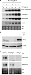
Silica nanoparticles promote an osteoblastic gene differentiation program. (A) NP1 dose-dependently induces expression of the characteristic osteoblastic gene products bone sialoprotein, osteocalcin, and osteopontin in MC3T3 cells, quantitated by northern blot. (B) Western blot of NP1 (50 µg/mL for 18 hr) stimulated expression of Runx2. TNFα (10 ng/ml), a known inhibitor of Runx2, was added as a control. (C) Osteocalcin and osteopontin are selectively upregulated by NP1 in pre-osteoblasts (Northern blot). Data representative of two independent experiments.
Figure 4
Figure 4
Silica Nanoparticles dose-dependently suppress NF-κB activation in MC3T3 cells and RAW264.7 cells. NP1 dose-dependently suppresses basal (A) and (B) RANKL-induced NF-κB activation in RAW264.7 cells, and (C) TNFα (10 ng/ml) induced NF-κB activation in MC3T3 cells. Cell lines were transfected with an NF-κB-responsive luciferase reporter and luciferase activity quantitated 24 hr later. Data expressed as Relative Light Units (RLU). (D) NP1 fails to suppress TNFα-induced NF-κB in HEK293 cells. All data points represent the average + SD of 4 replicate wells and 3 or more independent experiments.
Figure 5
Figure 5
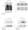
Silica nanoparticles antagonize NF-κB activation in osteoclast and osteoblast precursors. (A) MC3T3 cells were stimulated with TNFα (10 ng/mL) with or without NP1 (50 ng/mL) for 24 or 48 hr and nuclear extracts isolated for EMSA using radiolabeled NF-κB consensus probe. (B) MC3T3 cells were treated with TNFα and/or NP1 for 24 hr and whole cell extracts isolated for western blots. Blots were immunoprobed for NF-κB subunit p50 and its precursor p105. (C) RAW264.7 cells were treated with RANKL and/or NP1 for 24 hr and cytosolic and nuclear extracts isolated for Western blots. Blots were immunoprobed for NF-κB subunit p52. Actin and PCNA antibodies were used as loading controls for cytosol and nuclear extracts respectively. Densitometry scanning of bands and the actin/p52 or PCNA/p52 ratios are shown below the gels.
Figure 6
Figure 6
Silica nanoparticles enhance peak bone mineral density in mice in vivo. Female C57BL6 mice, 9 wks of age were injected intraperitoneally with NP1-MNP-PEG (50 mg/Kg) or vehicle, weekly for 6 weeks. BMD was quantitated at (A) the femur and (B) lumbar spine by DXA at baseline and at 2 week intervals up to 6 weeks and is presented as Mean ± SEM of percentage change from baseline, calculated for each mouse. For femurs, left and right femurs were averaged for each independent mouse. N= 9 mice per group. *p< 0.05, ** p< 0.01 or ***P< 0.001 by repeated measures ANOVA with Bonferroni multiple comparisons test.

References

    1. Riggs BL, Khosla S, Melton LJ., 3rd Sex steroids and the construction and conservation of the adult skeleton. Endocr Rev. 2002;23:279–302. - PubMed
    1. Cho M, Cho WS, Choi M, Kim SJ, Han BS, Kim SH, et al. The impact of size on tissue distribution and elimination by single intravenous injection of silica nanoparticles. Toxicol Lett. 2009;189:177–183. - PubMed
    1. Bartneck M, Keul HA, Zwadlo-Klarwasser G, Groll J. Phagocytosis independent extracellular nanoparticle clearance by human immune cells. Nano Lett. 2010;10:59–63. - PubMed
    1. Teitelbaum SL. Bone resorption by osteoclasts. Science. 2000;289:1504–1508. - PubMed
    1. Ducy P, Zhang R, Geoffroy V, Ridall AL, Karsenty G. Osf2/Cbfa1: a transcriptional activator of osteoblast differentiation. Cell. 1997;89:747–754. - PubMed

Publication types