

The intestinal microbiota are necessary for stressor-induced enhancement of splenic macrophage microbicidal activity
- PMID: 22100833
- PMCID: PMC3288745
- DOI: 10.1016/j.bbi.2011.11.002
The intestinal microbiota are necessary for stressor-induced enhancement of splenic macrophage microbicidal activity
Abstract
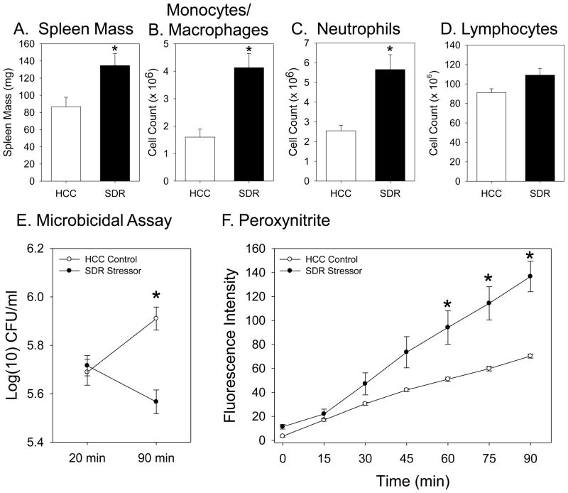
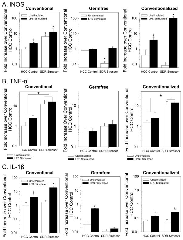
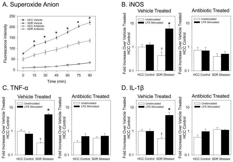
The indigenous microbiota impact mucosal, as well as systemic, immune responses, but whether the microbiota are involved in stressor-induced immunomodulation has not been thoroughly tested. A well characterized murine stressor, called social disruption (SDR), was used to study whether the microbiota are involved in stressor-induced enhancement of macrophage reactivity. Exposure to the SDR Stressor enhanced the ability of splenic macrophages to produce microbicidal mediators (e.g., inducible nitric oxide synthase (iNOS), superoxide anion, and peroxynitrite) and to kill target Escherichia coli. Exposure to the SDR Stressor also increased cytokine production by LPS-stimulated splenic macrophages. These effects, however, were impacted by the microbiota. Microbicidal activity and cytokine mRNA in splenic macrophages from Swiss Webster germfree mice that lack any commensal microbiota were not enhanced by exposure to the SDR Stressor. However, when germfree mice were conventionalized by colonizing them with microbiota from CD1 conventional donor mice, exposure to the SDR Stressor again increased microbicidal activity and cytokine mRNA. In follow-up experiments, immunocompetent conventional CD1 mice were treated with a cocktail of antibiotics to disrupt the intestinal microbiota. While exposure to the SDR Stressor-enhanced splenic macrophage microbicidal activity and cytokine production in vehicle-treated mice, treatment with antibiotics attenuated the SDR Stressor-induced increases in splenic macrophage reactivity. Treatment with antibiotics also prevented the stressor-induced increase in circulating levels of bacterial peptidoglycan, suggesting that translocation of microbiota-derived peptidoglycan into the body primes the innate immune system for enhanced activity. This study demonstrates that the microbiota play a crucial role in stressor-induced immunoenhancement.
Copyright © 2011 Elsevier Inc. All rights reserved.
Figures


Similar articles
-
Stressor-induced increase in microbicidal activity of splenic macrophages is dependent upon peroxynitrite production.Infect Immun. 2012 Oct;80(10):3429-37. doi: 10.1128/IAI.00714-12. Epub 2012 Jul 23. Infect Immun. 2012. PMID: 22825446 Free PMC article.
-
Exposure to a social stressor alters the structure of the intestinal microbiota: implications for stressor-induced immunomodulation.Brain Behav Immun. 2011 Mar;25(3):397-407. doi: 10.1016/j.bbi.2010.10.023. Epub 2010 Oct 30. Brain Behav Immun. 2011. PMID: 21040780 Free PMC article.
-
The contributing role of the intestinal microbiota in stressor-induced increases in susceptibility to enteric infection and systemic immunomodulation.Horm Behav. 2012 Aug;62(3):286-94. doi: 10.1016/j.yhbeh.2012.02.006. Epub 2012 Feb 15. Horm Behav. 2012. PMID: 22366706 Review.
-
Influence of stressor-induced nervous system activation on the intestinal microbiota and the importance for immunomodulation.Adv Exp Med Biol. 2014;817:255-76. doi: 10.1007/978-1-4939-0897-4_12. Adv Exp Med Biol. 2014. PMID: 24997038 Review.
-
Repeated social defeat increases the bactericidal activity of splenic macrophages through a Toll-like receptor-dependent pathway.Am J Physiol Regul Integr Comp Physiol. 2007 Sep;293(3):R1180-90. doi: 10.1152/ajpregu.00307.2007. Epub 2007 Jun 27. Am J Physiol Regul Integr Comp Physiol. 2007. PMID: 17596326
Cited by
-
The Role of the Intestinal Microbiome in Chronic Psychosocial Stress-Induced Pathologies in Male Mice.Front Behav Neurosci. 2018 Oct 26;12:252. doi: 10.3389/fnbeh.2018.00252. eCollection 2018. Front Behav Neurosci. 2018. PMID: 30464743 Free PMC article.
-
Impact of stressor exposure on the interplay between commensal microbiota and host inflammation.Gut Microbes. 2014 May-Jun;5(3):390-6. doi: 10.4161/gmic.28683. Epub 2014 Apr 1. Gut Microbes. 2014. PMID: 24690880 Free PMC article. Review.
-
Psychological stress creates an immune suppressive environment in the lung that increases susceptibility of aged mice to Mycobacterium tuberculosis infection.Front Cell Infect Microbiol. 2022 Sep 16;12:990402. doi: 10.3389/fcimb.2022.990402. eCollection 2022. Front Cell Infect Microbiol. 2022. PMID: 36189368 Free PMC article.
-
Stressor-induced increase in microbicidal activity of splenic macrophages is dependent upon peroxynitrite production.Infect Immun. 2012 Oct;80(10):3429-37. doi: 10.1128/IAI.00714-12. Epub 2012 Jul 23. Infect Immun. 2012. PMID: 22825446 Free PMC article.
-
The role of the commensal microbiota in adaptive and maladaptive stressor-induced immunomodulation.Horm Behav. 2017 Feb;88:70-78. doi: 10.1016/j.yhbeh.2016.10.006. Epub 2016 Oct 17. Horm Behav. 2017. PMID: 27760302 Free PMC article. Review.
References
-
- Avitsur R, Kavelaars A, Heijnen C, Sheridan JF. Social stress and the regulation of tumor necrosis factor-alpha secretion. Brain Behav Immun. 2005;19:311–317. - PubMed
Publication types
MeSH terms
Substances
Grants and funding
LinkOut - more resources
Full Text Sources
Medical
Molecular Biology Databases
Miscellaneous
