. 2012 Jan;33(1):23-36.
doi: 10.1016/j.neuro.2011.11.002. Epub 2011 Nov 9.

Neonatal Bisphenol A exposure alters sexually dimorphic gene expression in the postnatal rat hypothalamus

Affiliations

Neonatal Bisphenol A exposure alters sexually dimorphic gene expression in the postnatal rat hypothalamus

Jinyan Cao et al. Neurotoxicology. 2012 Jan.

Abstract

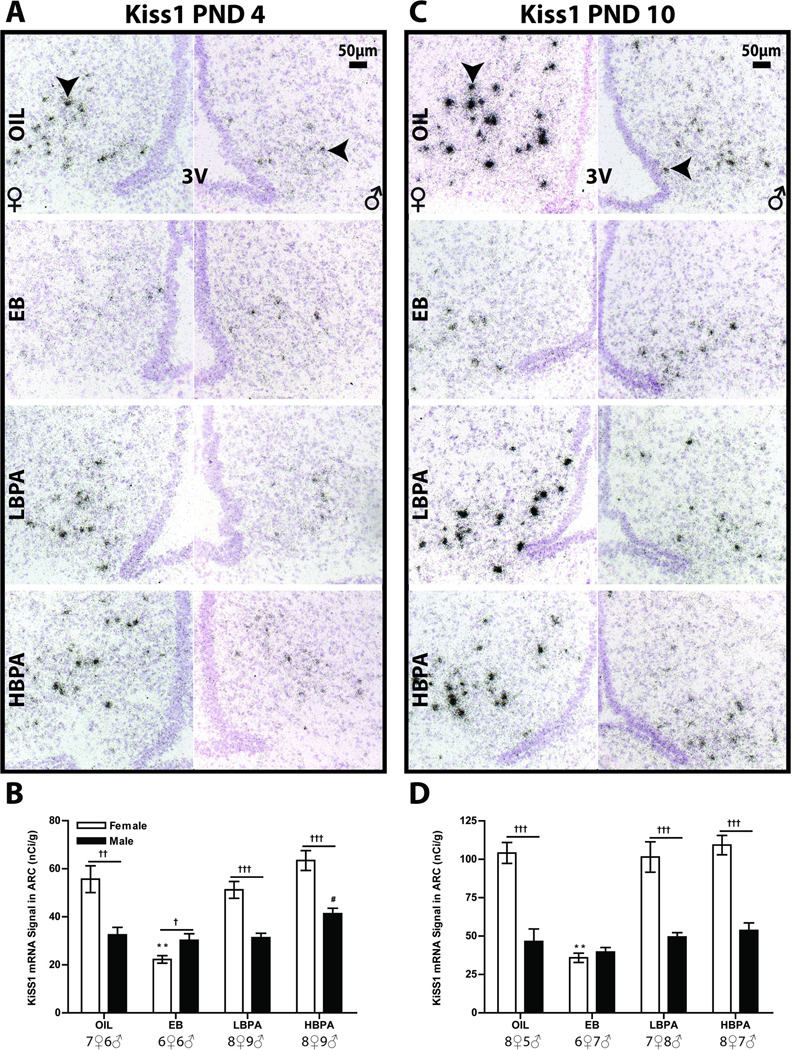
Developmental exposure to Bisphenol A (BPA), a component of polycarbonate and epoxy resins, has been purported to adversely impact reproductive function in female rodents. Because neonatal life is a critical window for the sexual dimorphic organization of the hypothalamic-pituitary-gonadal (HPG) axis, interference with this process could underlie compromised adult reproductive physiology. The goal of the present study was to determine if neonatal BPA exposure interferes with sex specific gene expression of estrogen receptor alpha (ERα), ER beta (ERβ) and kisspeptin (Kiss1) in the anterior and mediobasal hypothalamus. Long Evans (LE) neonatal rats were exposed to vehicle, 10μg estradiol benzoate (EB), 50mg/kg BPA or 50μg/kg BPA by subcutaneous injection daily from postnatal day 0 (PND 0) to PND 2. Gene expression was assessed by in situ hybridization on PNDs 4 and 10. Within the anterior hypothalamus ERα expression was augmented by BPA in PND 4 females, then fell to male-typical levels by PND 10. ERβ expression was not altered by BPA on PND 4, but significantly decreased or eliminated in both sexes by PND 10. Kiss1 expression was diminished by BPA in the anterior hypothalamus, especially in females. There were no significant impacts of BPA in the mediobasal hypothalamus. Collectively, BPA effects did not mirror those of EB. The results show that neonatal hypothalamic ER and Kiss1 expression is sensitive to BPA exposure. This disruption may alter sexually dimorphic hypothalamic organization and underlie adult reproductive deficiencies. Additionally, the discordant effects of EB and BPA indicate that BPA likely disrupts hypothalamic organization by a mechanism other than simply acting as an estrogen mimic.


Figures

Figure 1
Figure 1
Representative autoradiographs of ERα signal in the PND 4 AVPV (upper panels in A) and MPOA (lower panels in A), and in the PND 10 AVPV (upper panels in D) and MPOA (lower panels in D) after vehicle (OIL), EB, LBPA and HBPA treatments (from left to right in both A and D). Optical density analysis of ERα expression in the PND 4 AVPV (B) and MPOA (C), and in the PND 10 AVPV (E) and MPOA (F). ERα expression was significantly upregulated in the PND 4 HPBA females (B,C) then reduced to male-typical levels by PND 10 in both the AVPV (E) and the MPOA (F). LBPA had no effect in either sex on PND 4 but resulted in lower AVPV ERα levels in females by PND 10 (E). Significant differences in expression compared to vehicle are represented by *p ≤ 0.05, **p ≤ 0.01for the females, and #p ≤ 0.05, ##p ≤ 0.01 for the males. Significant sex differences in expression are represented by p ≤ 0.05, ††p ≤ 0.01, †††p ≤ 0.001. The graphs depict mean ± SEM and the sample size is provided at the bottom. (3V = third ventricle; AC = anterior commissure; scale bar = 500 µm)
Figure 2
Figure 2
Autoradiographs depicting ERα signal in the caudal VMNvl and ARC on PND 4 (A) and in the rostral VMNvl and ARC on PND 10 (F). Expression was sexually dimorphic within all regions examined and relatively unaltered by BPA exposure. The graphs depict mean ± SEM on PND 4 (B–E) and PND 10 (G–J) for rVMNvl, cVMNvl, rARC and cARC respectively with the sample size indicated at the bottom. Significant differences in expression compared to vehicle are represented by **p ≤ 0.01for the females, and #p ≤ 0.05, for the males. Significant sex differences in expression are represented by p ≤ 0.05, ††p ≤ 0.01, †††p ≤ 0.001. (3V = third ventricle; scale bar = 500 µm)
Figure 3
Figure 3
ERβ mRNA expression in the AVPV and MPOA of PND 4 (A) and PND 10 (D) rats following vehicle (OIL), EB, LBPA and HBPA treatment (A and D, from left to right). Expression was not sexually dimorphic on PND 4 and unaltered by exposure (B,C) but by PND 10, ERβ expression was depleted in the AVPV of both sexes (E) and significantly decreased in the female MPOA, thereby eliminating the sex difference (F). Significant differences in expression compared to vehicle are represented by *p ≤ 0.05, **p ≤ 0.01for the females, and #p ≤ 0.05, for the males. Significant sex differences in expression are represented by ††p ≤ 0.01, †††p ≤ 0.001. The graphs depict mean ± SEM and the sample size is provided at the bottom. (3V = third ventricle; AC = anterior commissure; scale bar = 500 µm)
Figure 4
Figure 4
Representative photomicrographs depicting ERβ mRNA labeling in the caudal VMNvl on PNDs 4 (A) and PND 10 (D). As expected, expression was absent in the ARC. In females, EB exposure reduced ERβ expression to male-typical levels throughout the VMNvl (B,C,E,F) while BPA had no appreciable effect with the exception of a slight elevation in the cVMNvl of the PND 10 males (F). Significant differences in expression compared to vehicle are represented by **p ≤ 0.01 for the females, and #p ≤ 0.05, ##p ≤ 0.01 for the males. Significant sex differences in expression are represented by ††p ≤ 0.01, †††p ≤ 0.001. The graphs depict mean ± SEM and the sample size is provided at the bottom. (3V = third ventricle; scale bar = 500 µm)
Figure 5
Figure 5
Hemotoxylin counterstained sections depicting silver grain labeling (black arrows) for Kiss1 in the PND 10 mid-level RP3V (A). Silver grain deposition was discrete and sexually dimorphic in the vehicle treated controls (A). The percentage of individuals with detectable clusters was nearly identical in the control groups but significantly lower or absent in the females exposed to EB or BPA an in the males exposed to EB or the low dose of BPA (n = 6–8 per sex per group) (B). Within the individuals displaying clusters, the mean number of clusters was sexually dimorphic in the control group but appreciably lower in females exposed to EB or BPA (C). No statistical analysis was performed because of the limited sample size. (scale bar = 50 µm)
Figure 6
Figure 6
Representative photomicrographs (A and C) depicting ARC Kiss1 silver grain clusters (black arrows) on PNDs 4 (A) and 10 (C). ARC expression levels were quantified from the film autoradiographs (not shown). In females, Kiss1 mRNA levels were significantly reduced in the EB exposure females on PNDs 4 (B) and 10 (D) and the sex difference was reversed or eliminated. BPA had no effect in females but Kiss1 levels were moderately upregulated in the HBPA males on PND 4 (B). Significant differences in expression compared to vehicle are represented by **p ≤ 0.01 for the females, and #p ≤ 0.05 for the males. Significant sex differences in expression are represented by p ≤ 0.05, ††p ≤ 0.01, †††p ≤ 0.001. The graphs depict mean ± SEM and the sample size is provided at the bottom. (3V = third ventricle; scale bar = 50 µm)

Similar articles

Cited by

References

    1. Adewale HB, Jefferson WN, Newbold RR, Patisaul HB. Neonatal Bisphenol-A Exposure Alters Rat Reproductive Development and Ovarian Morphology Without Impairing Activation of Gonadotropin Releasing Hormone Neurons. Biol Reprod. 2009 - PMC - PubMed
    1. Adewale HB, Todd KL, Mickens JA, Patisaul HB. The impact of neonatal bisphenol--a exposure on sexually dimorphic hypothalamic nuclei in the female rat. Neurotoxicology. 2011;32:38–49. - PMC - PubMed
    1. Aihara M, Hayashi S. Induction of persistent diestrus followed by persistent estrus is indicative of delayed maturation of tonic gonadotropin-releasing systems in rats. Biol Reprod. 1989;40:96–101. - PubMed
    1. Amateau SK, Alt JJ, Stamps CL, McCarthy MM. Brain estradiol content in newborn rats: sex differences, regional heterogeneity, and possible de novo synthesis by the female telencephalon. Endocrinology. 2004;145:2906–2917. - PubMed
    1. Andersen HR, Andersson AM, Arnold SF, Autrup H, Barfoed M, Beresford NA, et al. Comparison of short-term estrogenicity tests for identification of hormone-disrupting chemicals. Environmental health perspectives. 1999;107 Suppl 1:89–108. - PMC - PubMed

Publication types

MeSH terms

LinkOut - more resources