

Plasticity of serotonergic innervation of the inferior colliculus in mice following acoustic trauma
- PMID: 22101024
- PMCID: PMC3349240
- DOI: 10.1016/j.heares.2011.11.004
Plasticity of serotonergic innervation of the inferior colliculus in mice following acoustic trauma
Abstract
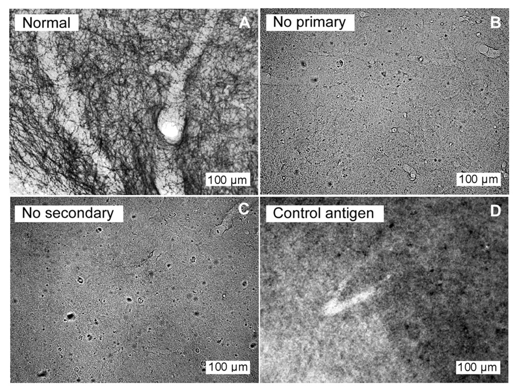
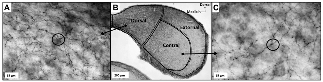
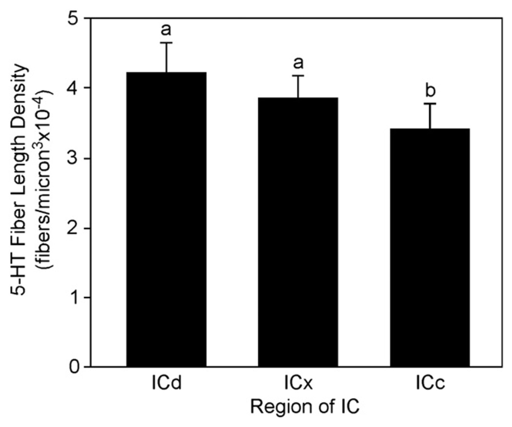
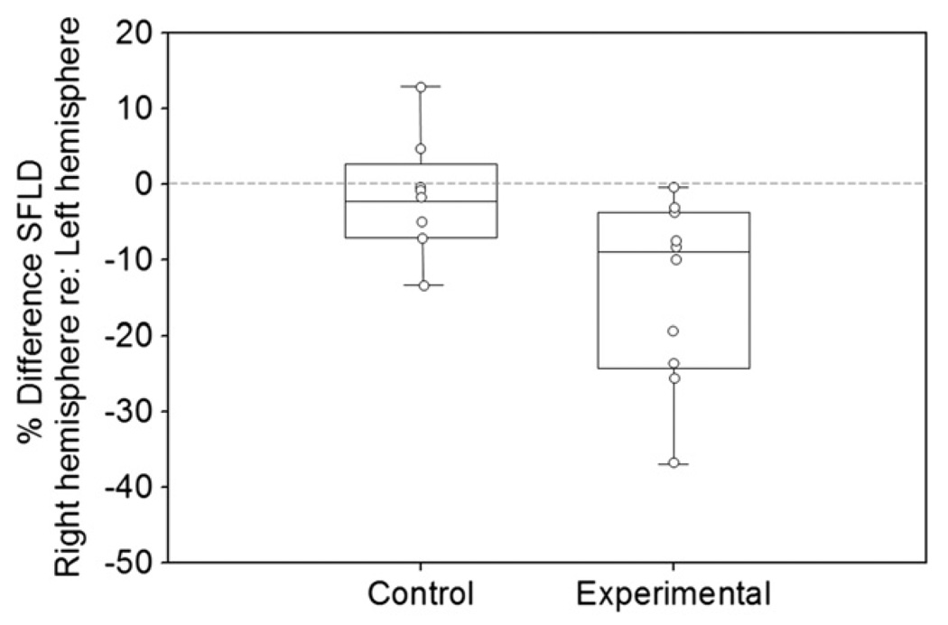
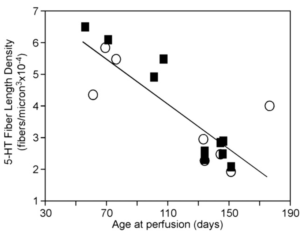
Acoustic trauma often results in permanent damage to the cochlea, triggering changes in processing within central auditory structures such as the inferior colliculus (IC). The serotonergic neuromodulatory system, present in the IC, is responsive to chronic changes in the activity of sensory systems. The current study investigated whether the density of serotonergic innervation in the IC is changed following acoustic trauma. The trauma stimulus consisted of an 8 kHz pure tone presented at a level of 113 dB SPL for six consecutive hours to anesthetized CBA/J mice. Following a minimum recovery period of three weeks, serotonergic fibers were visualized via histochemical techniques targeting the serotonin reuptake transporter (SERT) and quantified using stereologic probes. SERT-positive fiber densities were then compared between the traumatized and protected hemispheres of unilaterally traumatized subjects and those of controls. A significant effect of acoustic trauma was found between the hemispheres of unilaterally traumatized subjects such that the IC contralateral to the ear of exposure contained a lower density of SERT-positive fibers than the IC ipsilateral to acoustic trauma. No significant difference in density was found between the hemispheres of control subjects. Additional dimensions of variability in serotonergic fibers were seen among subdivisions of the IC and with age. The central IC had a slightly but significantly lowered density of serotonergic fibers than other subdivisions of the IC, and serotonergic fibers also declined with age. Overall, the results indicate that acoustic trauma is capable of producing modest but significant decreases in the density of serotonergic fibers innervating the IC.
Copyright © 2011 Elsevier B.V. All rights reserved.
Figures
Similar articles
-
Acoustic trauma triggers upregulation of serotonin receptor genes.Hear Res. 2014 Sep;315:40-8. doi: 10.1016/j.heares.2014.06.004. Epub 2014 Jul 2. Hear Res. 2014. PMID: 24997228 Free PMC article.
-
Social isolation reduces serotonergic fiber density in the inferior colliculus of female, but not male, mice.Brain Res. 2018 Sep 1;1694:94-103. doi: 10.1016/j.brainres.2018.05.010. Epub 2018 May 12. Brain Res. 2018. PMID: 29763575
-
Socially induced serotonergic fluctuations in the male auditory midbrain correlate with female behavior during courtship.J Neurophysiol. 2016 Apr;115(4):1786-96. doi: 10.1152/jn.00742.2015. Epub 2016 Jan 20. J Neurophysiol. 2016. PMID: 26792882 Free PMC article.
-
Functional reorganization in chinchilla inferior colliculus associated with chronic and acute cochlear damage.Hear Res. 2002 Jun;168(1-2):238-49. doi: 10.1016/s0378-5955(02)00360-x. Hear Res. 2002. PMID: 12117524 Review.
-
Hearing loss-related altered neuronal activity in the inferior colliculus.Hear Res. 2024 Aug;449:109033. doi: 10.1016/j.heares.2024.109033. Epub 2024 May 20. Hear Res. 2024. PMID: 38797036 Review.
Cited by
-
Resveratrol prevents hearing loss and a subregion specific- reduction of serotonin reuptake transporter induced by noise exposure in the central auditory system.Front Neurosci. 2023 Mar 24;17:1134153. doi: 10.3389/fnins.2023.1134153. eCollection 2023. Front Neurosci. 2023. PMID: 37034161 Free PMC article.
-
Listening to your partner: serotonin increases male responsiveness to female vocal signals in mice.Front Hum Neurosci. 2024 Jan 24;17:1304653. doi: 10.3389/fnhum.2023.1304653. eCollection 2023. Front Hum Neurosci. 2024. PMID: 38328678 Free PMC article.
-
Silence, Solitude, and Serotonin: Neural Mechanisms Linking Hearing Loss and Social Isolation.Brain Sci. 2020 Jun 12;10(6):367. doi: 10.3390/brainsci10060367. Brain Sci. 2020. PMID: 32545607 Free PMC article. Review.
-
Behavioral Animal Model of the Emotional Response to Tinnitus and Hearing Loss.J Assoc Res Otolaryngol. 2018 Feb;19(1):67-81. doi: 10.1007/s10162-017-0642-8. Epub 2017 Oct 18. J Assoc Res Otolaryngol. 2018. PMID: 29047013 Free PMC article.
-
From behavioral context to receptors: serotonergic modulatory pathways in the IC.Front Neural Circuits. 2012 Sep 6;6:58. doi: 10.3389/fncir.2012.00058. eCollection 2012. Front Neural Circuits. 2012. PMID: 22973195 Free PMC article.
References
-
- Abbott SD, Hughes LF, Bauer CA, Salvi R, Caspary DM. Detection of glutamate decarboxylase isoforms in rat inferior colliculus following acoustic exposure. Neuroscience. 1999;93(4):1375–1381. - PubMed
-
- Baroncelli L, Sale A, Viegi A, Vetencourt JFM, De Pasquale R, Baldini S, Maffei L. Experience-dependent reactivation of ocular dominance plasticity in the adult visual cortex. Exp. Neurol. 2010;226(1):100–109. - PubMed
-
- Bartels H, Staal MJ, Albers FWJ. Tinnitus and neural plasticity of the brain. Otol. Neurotol. 2007;28:178–184. - PubMed
-
- Basta D, Ernst A. Erratum to “Noise-induced changes of neuronal spontaneous activity in mice inferior colliculus brain slices. Neurosci. Lett. 2005;374:74–79. - PubMed
Publication types
MeSH terms
Substances
Grants and funding
LinkOut - more resources
Full Text Sources
Medical
Miscellaneous
