

Rapid measurement of antituberculosis drug activity in vitro and in macrophages using bioluminescence
- PMID: 22101217
- PMCID: PMC3254196
- DOI: 10.1093/jac/dkr472
Rapid measurement of antituberculosis drug activity in vitro and in macrophages using bioluminescence
Abstract
Objectives: Tuberculosis drug development is hampered by the slow growth of Mycobacterium tuberculosis. Bioluminescence, light produced by an enzymatic reaction, constitutes a rapid and highly sensitive measurement of cell metabolic function that can be used as an indirect marker of cell viability in drug screening assays. The aim of this work was to validate and standardize the use of luminescent M. tuberculosis strains to test the activity of antibacterial drugs in vitro and inside macrophages in a 96-well format.
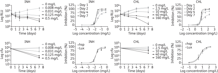
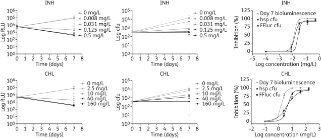
Methods: We have used strains that express the bacterial lux operon and therefore do not require exogenous substrate to produce light, as well as strains expressing the firefly luciferase that need luciferin substrate. Results were compared with those obtained using the resazurin reduction assay and cfu plating.
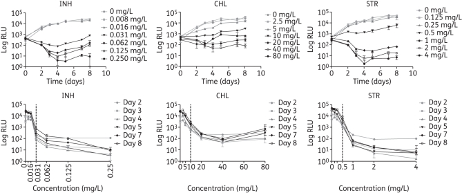
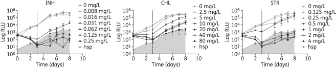
Results: Using bioluminescence we were able to reduce the time required to measure the MIC and bactericidal concentrations of antimicrobials to just 3 and 6 days, respectively. Furthermore, antibacterial activity against intracellular mycobacteria was detected within 2 days post-infection. Results were comparable to those obtained by conventional methods.
Conclusions: We have developed a simple and rapid method for screening antimycobacterial drugs in culture and in macrophages. The use of autoluminescent bacteria also facilitates the determination of growth and inhibition kinetics. The method is cost-effective, can easily be adapted to a larger scale and is amenable to automation. Current efforts are directed towards applying this technology to drug screening in vivo.
Figures
References
-
- WHO. Global Tuberculosis Control 2010. Geneva: WHO Press; 2010.
-
- Orme I. Search for new drugs for treatment of tuberculosis. Antimicrob Agents Chemother. 2001;45:1943–6. doi:10.1128/AAC.45.7.1943-1946.2001. - DOI - PMC - PubMed
-
- Primm TP, Franzblau SG. Recent advances in methodologies for the discovery of antimycobacterial drugs. Curr Bioact Compd. 2007;3:201–8. doi:10.2174/157340707781695550. - DOI
-
- Caviedes L, Delgado J, Gilman RH. Tetrazolium microplate assay as a rapid and inexpensive colorimetric method for determination of antibiotic susceptibility of Mycobacterium tuberculosis. J Clin Microbiol. 2002;40:1873–4. doi:10.1128/JCM.40.5.1873-1874.2002. - DOI - PMC - PubMed
