Review
. 2012 Jan 28;41(4):1118-27.
doi: 10.1039/c1dt11535a. Epub 2011 Nov 18.

Vanadium nitrogenase: a two-hit wonder?

Affiliations
Review

Vanadium nitrogenase: a two-hit wonder?

Yilin Hu et al. Dalton Trans. .

Abstract

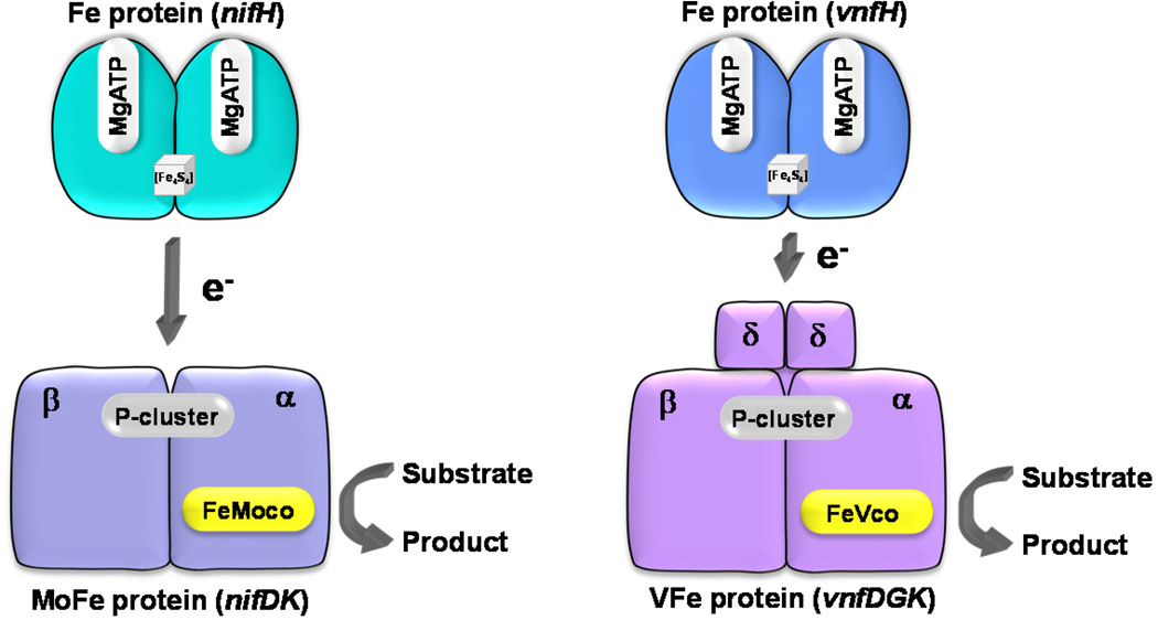
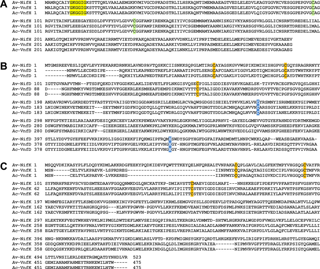
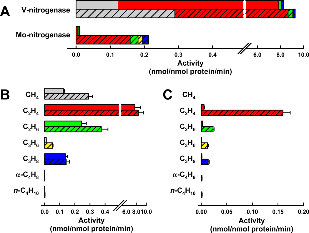
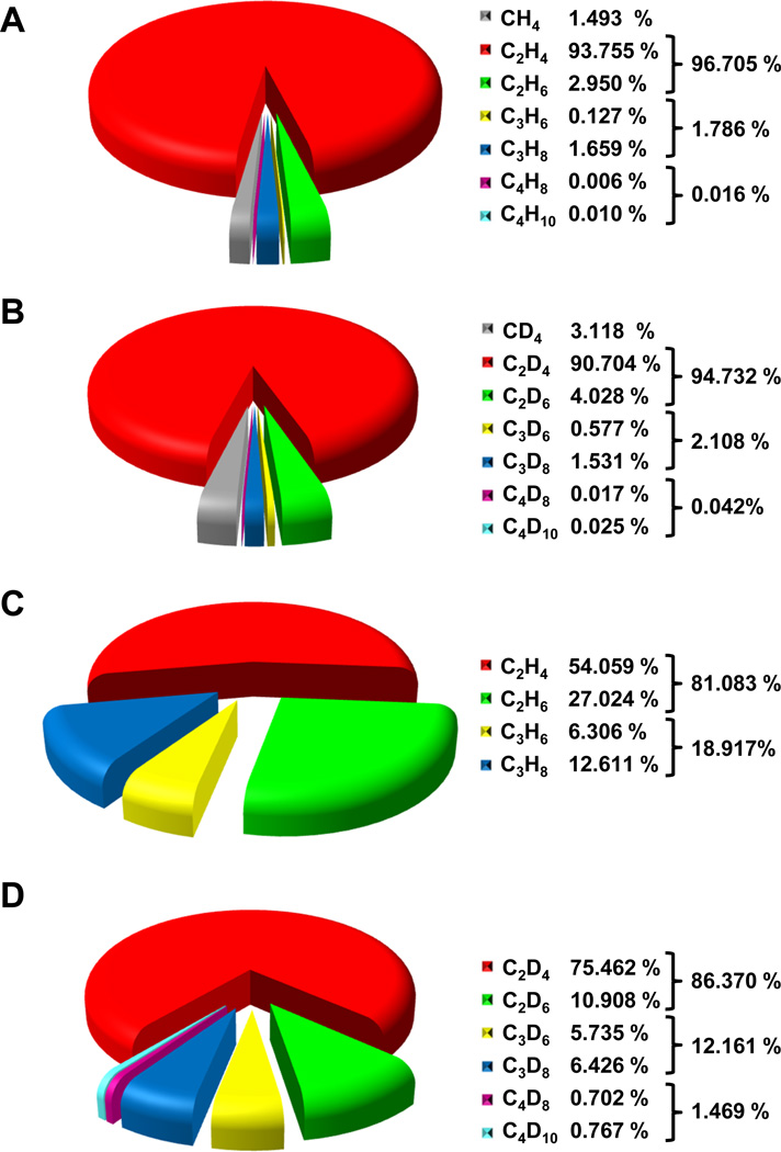
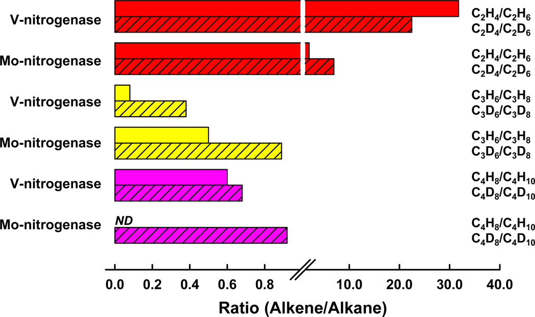
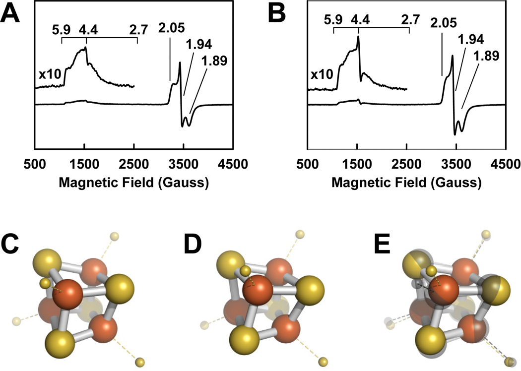
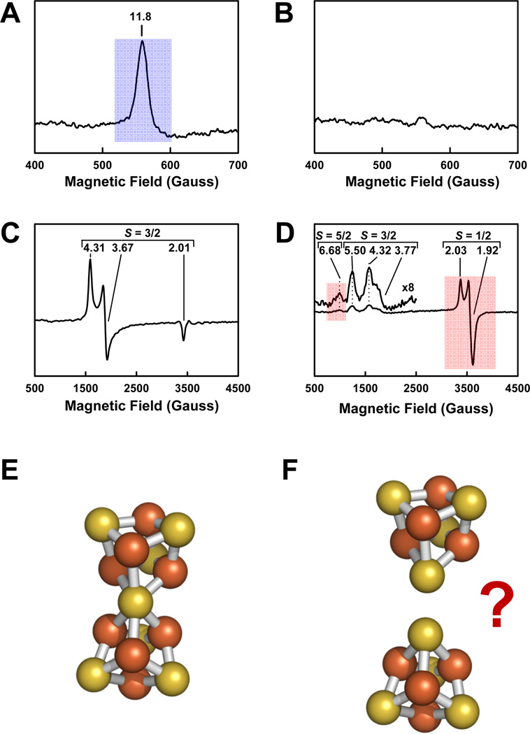
Nitrogenase catalyzes the biological conversion of atmospheric dinitrogen to bioavailable ammonia. The molybdenum (Mo)- and vanadium (V)-dependent nitrogenases are two homologous members of this metalloenzyme family. However, despite their similarities in structure and function, the characterization of V-nitrogenase has taken a much longer and more winding path than that of its Mo-counterpart. From the initial discovery of this nitrogen-fixing system, to the recent finding of its CO-reducing capacity, V-nitrogenase has proven to be a two-hit wonder in the over-a-century-long research of nitrogen fixation. This perspective provides a brief account of the catalytic function and structural basis of V-nitrogenase, as well as a short discussion of the theoretical and practical potentials of this unique metalloenzyme.


Figures

Fig. 1
Fig. 1. X-ray crystal structures of the component proteins and the complex of Mo-nitrogenase
(A, B) The γ2-dimeric Fe protein (A) and the α2β2-tetrameric MoFe protein (B) of A. vinelandii. (C, D) Half of the ADP•AlF4-stabilized Fe protein/MoFe protein complex (C) and the relative positions of components that are involved in the electron flow during catalysis (D). The two subunits of Fe protein are colored yellow and orange, and the α- and β-subunits of MoFe protein are colored red and blue, respectively. The positions of [Fe4S4] cluster, P-cluster and FeMoco are indicated. All clusters and ADP•AlF4 are shown as space-filling models, with the atoms colored as follows: Fe, orange; S, yellow; Mo, cyan; O, red; C, gray; N, dark blue; X (C, N or O), light gray; Mg, green; Al, beige; F, light blue. PYMOL was used to create the figure (PDB entries 1NIP, 1M1N and 1N2C).
Fig. 2
Fig. 2. Catalytic units of Mo- and V-nitrogenases
Schematic presentations of the catalytic unit of Mo-nitrogenase (left), which consists of the γ2-dimeric Fe protein (encoded by nifH) and one half of the α2β2-tetrameric MoFe protein (encoded by nifDK); and the catalytic unit of V-nitrogenase (right), which consists of the γ2-dimeric Fe protein (encoded by vnfH) and one half of the α2β2δ4-octameric VFe protein (encoded by vnfDGK). It is hypothesized that, during catalysis, the Fe protein forms a functional complex with the MoFe or VFe protein, in which electrons are sequentially transferred from the [Fe4S4] cluster (of the Fe protein), through the P-cluster, to the FeMoco (of MoFe protein) or the FeVco (of VFe protein), where substrate reduction eventually occurs.
Fig. 3
Fig. 3. Comparison between the primary sequences of Mo- and V-nitrogenases
(A) Sequence alignment of the Fe proteins of A. vinelandii Mo-nitrogenase (Av-NifH), A. vinelandii V-nitrogenase (Av-VnfH) and A. chroococcum V-nitrogenase (Ac-VnfH). The two conserved Cys residues, which serve as the [Fe4S4] cluster ligands, are highlighted in green; and the Gly-X-Gly-X-X-Gly consensus nucleotide-binding motifs are highlighted in yellow. (B) Sequence alignment of the α-subunits of A. vinelandii MoFe protein (Av-NifD), A. vinelandii VFe protein (Av-VnfD) and A. chroococcum VFe protein (Ac-VnfD). (C) Sequence alignment of the β-subunits of A. vinelandii MoFe protein (Av-NifK), A. vinelandii VFe protein (Av-VnfK) and A. chroococcum VFe protein (Ac-VnfK). The VFe protein contains an additional δ-subunit (Av- or Ac-VnfG), which is absent from the MoFe protein. The six conserved Cys residues, which serve as the P-cluster ligands, are highlighted in orange; and the conserved Cys and His residues, which serve as the FeMoco ligands, are highlighted in blue.
Fig. 4
Fig. 4. Specific activities of CO reduction by V- and Mo-nitrogenases
(A) Total activities of product formation by the two nitrogenases in the presence of H2O (solid) or D2O (striped). (B, C) Specific activities of individual product formation by V (B)- or Mo (C)-nitrogenase.
Fig. 5
Fig. 5. Product distributions of CO reduction by V- and Mo-nitrogenases
(A, B) Product profile of V-nitrogenase in the presence of H2O (A) or D2O (B). (C, D) Product profile of Mo-nitrogenase in the presence of H2O (C) or D2O (D). The total amounts of hydrocarbons detected in V (A, B)- and Mo (C, D)-nitrogenase-catalyzed reactions were set as 100%, respectively, and the percentages of individual products in these reactions were determined accordingly for each nitrogenase.
Fig. 6
Fig. 6. Ratios between alkenes and alkanes in reactions catalyzed by V- and Mo-nitrogenases
Shown are alkene/alkane ratios of the two (red)-, three (yellow)- and four (pink)-carbon products in the presence of H2O (solid) or D2O (striped).
Fig. 7
Fig. 7. [Fe4S4] clusters of the Fe proteins of Mo-nitrogenase (NifH) and V-nitrogenase (VnfH)
(A, B) Dithionite-reduced EPR spectra of NifH (A) and VnfH (B). (C–E) XAS/EXAFS-derived structures of the [Fe4S4] clusters in NifH (C) and VnfH (D), and the structural overlay of the two [Fe4S4] clusters (E). The clusters are shown as ball-and-stick models, with the atoms colored as those in Fig. 1. PYMOL was used to create the figure.
Fig. 8
Fig. 8. P-clusters of the MoFe and VFe proteins
(A, B) Indigo disulfonate (IDS)-oxidized EPR spectra of MoFe (A) and VFe (B) proteins. (C, D) Dithionite-reduced EPR spectra of MoFe (C) and VFe (D) proteins. The EPR feature associated with the P-cluster in MoFe protein is highlighted in blue shade, and those associated with the P-cluster in VFe protein are highlighted in red shade. The g values are indicated. (E, F) Crystal structure of the P-cluster in MoFe protein (E) and proposed structure of the P-cluster in VFe protein (F). The clusters are shown as ball-and-stick models, with the atoms colored as those in Fig. 1. PYMOL was used to create the figure.
Fig. 9
Fig. 9. Cofactors of the MoFe and VFe proteins
(A, C) Dithionite-reduced EPR spectra of MoFe protein (A) and NMF-extracted FeMoco (C). (B, D) Dithionite-reduced EPR spectra of VFe protein (B) and NMF-extracted FeVco (D). The corresponding EPR features in the protein-bound and NMF-extracted states are highlighted in blue shade for the FeMoco (A, C) and in red shade for the FeVco (B, D). The g values are indicated. (E, F) The XAS/EXAFS-derived structures of NMF-extracted FeMoco (E) and FeVco (F). The clusters are shown as ball-and-stick models, whereas the NMF molecules are represented by sticks, with the atoms colored as those in Fig. 1. PYMOL was used to create the figure.
Fig. 10
Fig. 10. Specific activities of the “standard” substrate reduction by reconstituted and native nitrogenases
Specific activities of FeVco (A)- and FeMoco (B)-reconstituted MoFe proteins and native VFe (C) and MoFe (D) proteins in H2 formation under Ar (black), C2H4 (red) and C2H6 (green) formation under C2H2, and NH3 (yellow) and H2 (blue) formation under N2. ①, characteristic catalytic features of V-nitrogenase; ②, characteristic catalytic features of Mo-nitrogenase.

Similar articles

Cited by

References

    1. Burgess BK, Lowe DJ. Chem. Rev. 1996;96:2983–3012. - PubMed
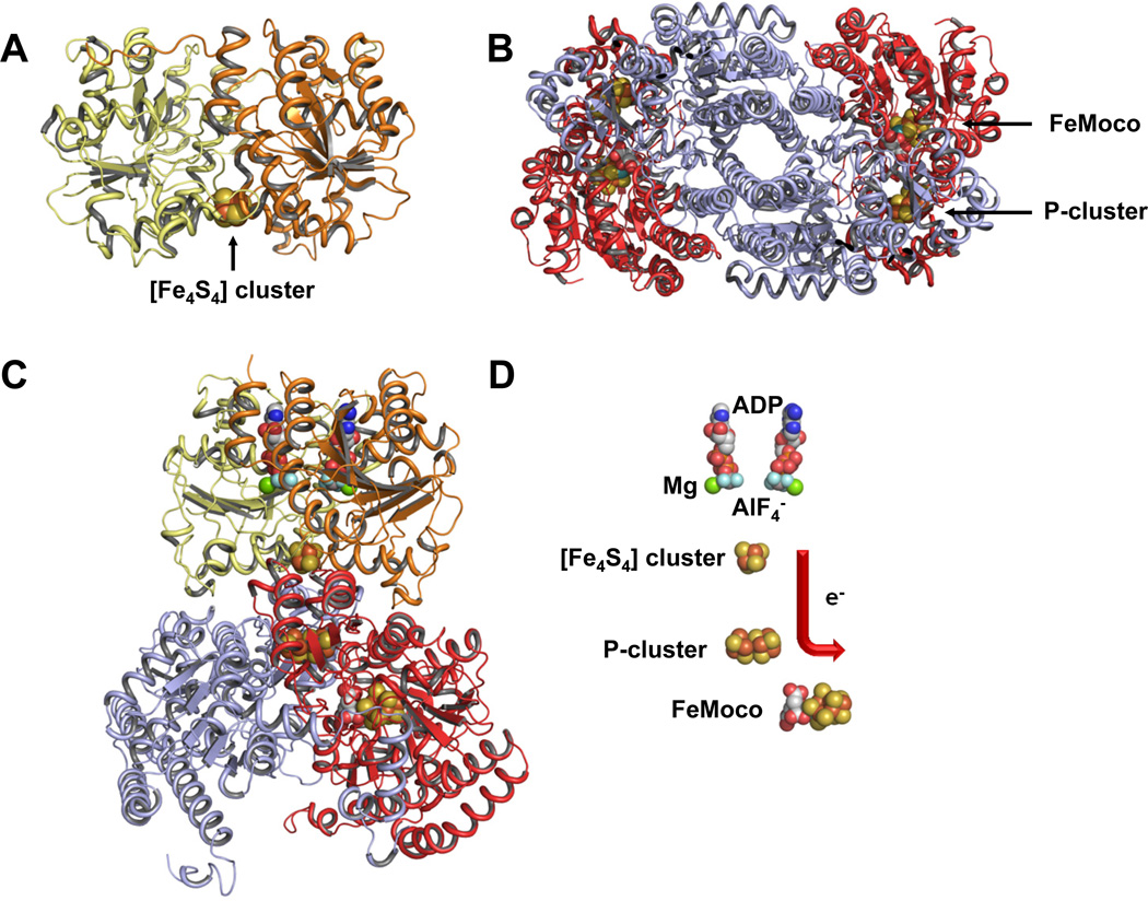
    1. Georgiadis MM, Komiya H, Chakrabarti P, Woo D, Kornuc JJ, Rees DC. Science. 1992;257:1653–1659. - PubMed
    1. Einsle O, Tezcan FA, Andrade SL, Schmid B, Yoshida M, Howard JB, Rees DC. Science. 2002;297:1696–1700. - PubMed
    1. Schindelin H, Kisker C, Schlessman JL, Howard JB, Rees DC. Nature. 1997;387:370–376. - PubMed
    1. Bishop PE, Premakumar R. In: Biological Nitorgen Fixation. Stacey G, Burris RH, Evans HJ, editors. New York: Chapman and Hall; 1992. pp. 736–762.

Publication types