. 2012 Feb 1;302(3):H724-32.
doi: 10.1152/ajpheart.00739.2011. Epub 2011 Nov 18.

Novel role of p66Shc in ROS-dependent VEGF signaling and angiogenesis in endothelial cells

Affiliations

Novel role of p66Shc in ROS-dependent VEGF signaling and angiogenesis in endothelial cells

Jin Oshikawa et al. Am J Physiol Heart Circ Physiol. .

Abstract

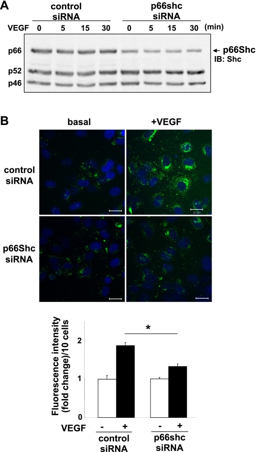
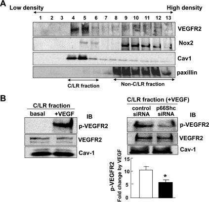
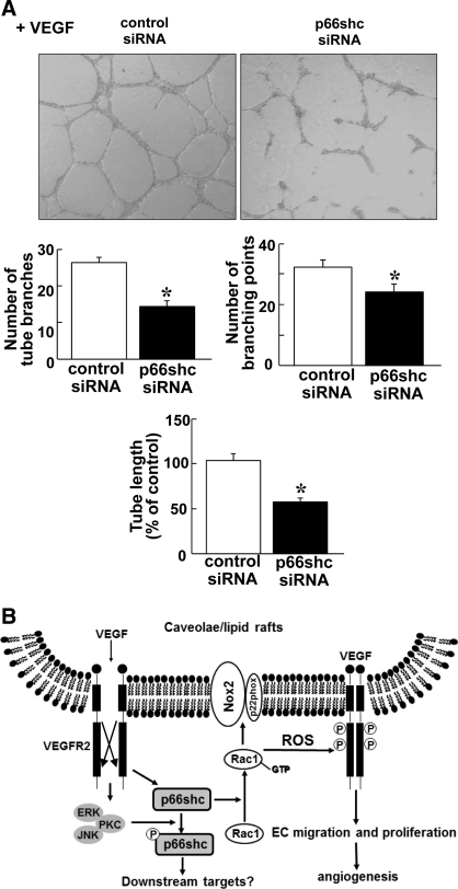
p66Shc, a longevity adaptor protein, is demonstrated as a key regulator of reactive oxygen species (ROS) metabolism involved in aging and cardiovascular diseases. Vascular endothelial growth factor (VEGF) stimulates endothelial cell (EC) migration and proliferation primarily through the VEGF receptor-2 (VEGFR2). We have shown that ROS derived from Rac1-dependent NADPH oxidase are involved in VEGFR2 autophosphorylation and angiogenic-related responses in ECs. However, a role of p66Shc in VEGF signaling and physiological responses in ECs is unknown. Here we show that VEGF promotes p66Shc phosphorylation at Ser36 through the JNK/ERK or PKC pathway as well as Rac1 binding to a nonphosphorylated form of p66Shc in ECs. Depletion of endogenous p66Shc with short interfering RNA inhibits VEGF-induced Rac1 activity and ROS production. Fractionation of caveolin-enriched lipid raft demonstrates that p66Shc plays a critical role in VEGFR2 phosphorylation in caveolae/lipid rafts as well as downstream p38MAP kinase activation. This in turn stimulates VEGF-induced EC migration, proliferation, and capillary-like tube formation. These studies uncover a novel role of p66Shc as a positive regulator for ROS-dependent VEGFR2 signaling linked to angiogenesis in ECs and suggest p66Shc as a potential therapeutic target for various angiogenesis-dependent diseases.


Figures

Fig. 1.
Fig. 1.
VEGF increases Ser36 phosphorylation of p66Shc through ERK, JNK, or PKC in endothelial cells (ECs). A: human umbilical vein ECs (HUVECs) were stimulated with VEGF (20 ng/ml) for indicated minutes, and lysates were immunoprecipitated (IP) with anti-Shc antibody (Ab) or normal IgG (negative control) and immunoblotted (IB) with anti-phospho-Ser36-p66Shc (pS36-p66Shc) Ab or Shc Ab. (n = 3). Bottom: averaged data, expressed as fold change over basal (means ± SE). *P < 0.05 vs. control. B: HUVECs were pretreated with 20 μM LY294002 (LY), 10 μM SB203580 (SB), 20 μM PD98059 (PD), 20 μM SP600125 (SP), or 10 μM GF109203X (GF) for 30 min and stimulated with VEGF (20 ng/ml) for 15 min. Lysates were measured for pS36-p66Shc (n = 5). *P < 0.05 vs. vehicle +VEGF.
Fig. 2.
Fig. 2.
p66Shc is involved in VEGF-induced reactive oxygen species (ROS) production in ECs. A: Western blots analysis for p66Shc in HUVECs after p66Shc short interfering (si)RNA treatment for 48 h. Lysates were immunoblotted with anti-total Shc Ab, and the bands of p66Shc are shown at 66 kDa. B, top: dichlorofluorescein (DCF) fluorescence with DAPI staining was measured by confocal microscopy in HUVECs transfected with control or p66Shc siRNAs with or without 20 ng/ml VEGF simulation for 5 min. B, bottom: averaged data of DCF fluorescence intensity (fold change)/10 cells (n = 3). *P < 0.05.
Fig. 3.
Fig. 3.
VEGF stimulates p66Shc association with Rac1 and its activation through p66Shc in ECs. A: HUVECs were stimulated with VEGF (20 ng/ml) for indicated minutes. Cell lysates were IP with anti-Rac1 or Shc Abs or normal IgG, and IB with anti-Shc or pS36-p66Shc Abs. Lysates without IP (No IP) were IB with anti-Rac1 Ab. A, bottom: averaged data for fold change in p66Shc-Rac1 association, expressed as -fold change over basal. *P < 0.05. B: Rac1 activities were measured in HUVECs transfected with control or p66Shc siRNAs with or without 20 ng/ml VEGF stimulation for 5 min (n = 3). *P < 0.05.
Fig. 4.
Fig. 4.
p66Shc is involved in VEGF-induced VEGFR2 autophosphorylation and p38MAPK activation in ECs. HUVECs were transfected with control or p66Shc siRNAs. A: cells were stimulated with VEGF (20 ng/ml) for 5 min, and lysates were IP with anti-VEGFR2 Ab and IB with anti-phospho-tyrosine (pTyr) Ab. Same lysates were IB with anti-VEGFR2 or Shc Abs (n = 4) *P < 0.05. B: cells were stimulated with VEGF for indicated minutes and lysates were IB with phospho-p38MAPK or phospho-ERK1/2 or p66Shc expression. *P < 0.05.
Fig. 5.
Fig. 5.
p66Shc is involved in VEGF-induced VEGFR2 autophosphorylation in caveolae/lipid rafts in ECs. A: sucrose gradient centrifugation was performed to isolate caveolae/lipid rafts (C/LR), equal volume of each fraction from top to bottom was IB with anti-VEGFR2, Nox2, caveolin-1, and paxillin (marker for non-C/LR) Abs. C/LR fraction were appeared in fractions 4–6. B, left: HUVECs were stimulated with or without VEGF (20 ng/ml) for 5 min, and followed by C/LR fractionation. Equal amounts of proteins from C/LR fraction were IB with anti-VEGFR2-pY1175, total VEGFR2, and caveolin-1 Abs (n = 3). B, right: HUVECs transfected with control or p66Shc siRNAs were stimulated with VEGF (20 ng/ml) for 5 min, and the C/LR fraction were IB with anti-VEGFR2-pY1175, total VEGFR2, and caveolin-1 Abs. B, bottom: averaged data, expressed as VEGF-induced fold change over basal in control siRNA-treated groups (means ± SE). *P < 0.05.
Fig. 6.
Fig. 6.
p66Shc is involved in VEGF-induced cell migration in ECs. HUVECs transfected with control or p66Shc siRNAs were used for measurement of EC migration using modified Boyden chamber assay (A) or wound scratch assay (B) as well as EC proliferation (C). A: cell migration stimulated with VEGF for 6 h was measured using modified Boyden chamber method. Bar graph represents averaged data, expressed as cell number counted per 10 fields (x 200) and fold change over that in unstimulated cells (control). *P < 0.05. B: confluent monolayer of HUVECs were wound scratched in the presence of VEGF (20 ng/ml) to stimulate the EC migration toward the wound area. Images were captured immediately (0 h) and 24 h after the wound scratch. C: cells were cultured in 0.2% FBS containing medium with or without 20 ng/ml VEGF for 48 h and the cell number was measured. *P < 0.05, for the fold increase induced by VEGF vs. vehicle alone (n = 3).
Fig. 7.
Fig. 7.
Role of p66Shc in capillary-like network formation in ECs and proposed model. A: HUVECs were seeded on Matrigel-coated plates in culture media containing VEGF for 6 h. Eight random fields per well were imaged, and representative pictures are shown (top). Averaged numbers of capillary tube branches, branching points, and tube length per field are shown (bottom; n = 3). P < 0.05. B: proposed model for novel role of p66Shc in ROS-dependent VEGFR2 signaling and angiogenesis in ECs. VEGF stimulation promotes pS36-pp66Shc formation through ERK/JNK/PKC as well as association of nonphosphorylated form of p66Shc with Rac1, a component of Nox2 NADPH oxidase in ECs. p66Shc is involved in VEGF-induced Rac1 activation and ROS production, thereby promoting VEGFR2 activation in caveolae/lipid rafts, which in turn stimulates angiogenic responses in ECs.

References

    1. Arany I, Faisal A, Nagamine Y, Safirstein RL. p66Shc inhibits pro-survival epidermal growth factor receptor/ERK signaling during severe oxidative stress in mouse renal proximal tubule cells. J Biol Chem 283: 6110–6117, 2008 - PubMed
    1. Babior BM. The NADPH oxidase of endothelial cells. IUBMB Life 50: 267–269, 2000 - PubMed
    1. Camici GG, Schiavoni M, Francia P, Bachschmid M, Martin-Padura I, Hersberger M, Tanner FC, Pelicci P, Volpe M, Anversa P, Luscher TF, Cosentino F. Genetic deletion of p66(Shc) adaptor protein prevents hyperglycemia-induced endothelial dysfunction and oxidative stress. Proc Natl Acad Sci USA 104: 5217–5222, 2007 - PMC - PubMed
    1. Carpi A, Menabo R, Kaludercic N, Pelicci P, Di Lisa F, Giorgio M. The cardioprotective effects elicited by p66(Shc) ablation demonstrate the crucial role of mitochondrial ROS formation in ischemia/reperfusion injury. Biochim Biophys Acta 1787: 774–780, 2009 - PubMed
    1. Chahdi A, Sorokin A. Endothelin-1 couples betaPix to p66Shc: role of betaPix in cell proliferation through FOXO3a phosphorylation and p27kip1 down-regulation independently of Akt. Mol Biol Cell 19: 2609–2619, 2008 - PMC - PubMed

Publication types

MeSH terms

Substances