Review
. 2012 Feb 1;302(3):H527-37.
doi: 10.1152/ajpheart.00676.2011. Epub 2011 Nov 18.

Central neural control of sympathetic nerve activity in heart failure following exercise training

Affiliations
Review

Central neural control of sympathetic nerve activity in heart failure following exercise training

Kaushik P Patel et al. Am J Physiol Heart Circ Physiol. .

Abstract

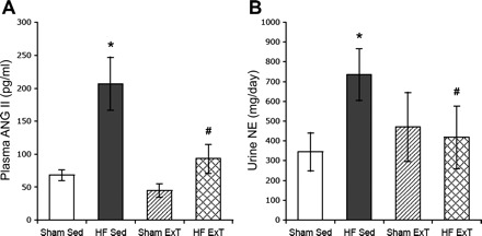
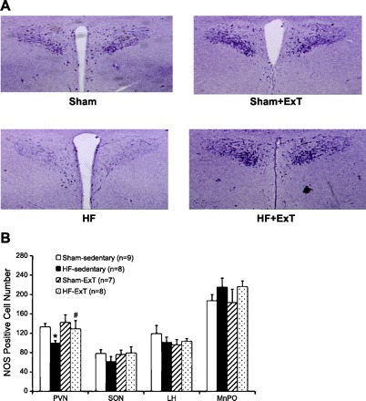
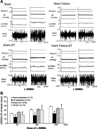
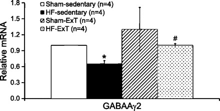
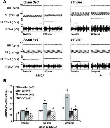
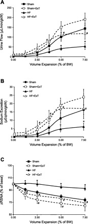
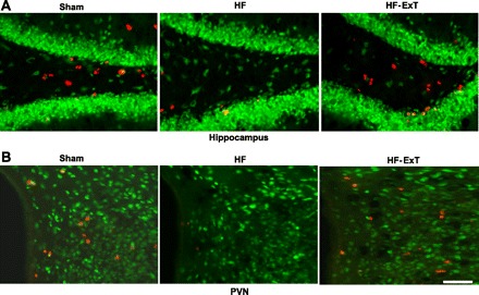
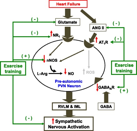
Typical characteristics of chronic congestive heart failure (HF) are increased sympathetic drive, altered autonomic reflexes, and altered body fluid regulation. These abnormalities lead to an increased risk of mortality, particularly in the late stage of chronic HF. Recent evidence suggests that central nervous system (CNS) mechanisms may be important in these abnormalities during HF. Exercise training (ExT) has emerged as a nonpharmacological therapeutic strategy substitute with significant benefit to patients with HF. Regular ExT improves functional capacity as well as quality of life and perhaps prognosis in chronic HF patients. The mechanism(s) by which ExT improves the clinical status of HF patients is not fully known. Recent studies have provided convincing evidence that ExT significantly alleviates the increased sympathetic drive, altered autonomic reflexes, and altered body fluid regulation in HF. This review describes and highlights the studies that examine various central pathways involved in autonomic outflow that are altered in HF and are improved following ExT. The increased sympathoexcitation is due to an imbalance between inhibitory and excitatory mechanisms within specific areas in the CNS such as the paraventricular nucleus (PVN) of the hypothalamus. Studies summarized here have revealed that ExT improves the altered inhibitory pathway utilizing nitric oxide and GABA mechanisms within the PVN in HF. ExT alleviates elevated sympathetic outflow in HF through normalization of excitatory glutamatergic and angiotensinergic mechanisms within the PVN. ExT also improves volume reflex function and thus fluid balance in HF. Preliminary observations also suggest that ExT induces structural neuroplasticity in the brain of rats with HF. We conclude that improvement of the enhanced CNS-mediated increase in sympathetic outflow, specifically to the kidneys related to fluid balance, contributes to the beneficial effects of ExT in HF.


Figures

Fig. 1.
Fig. 1.
Plasma angiotensin II (ANG II; A) and urinary norepinephrine (NE; B) in sham-sedentary (Sed); heart failure (HF)-Sed, sham-exercise-trained (ExT), and HF-ExT rats. Values are means ± SE. *P < 0.05, significantly different from the respective sham group. #P < 0.05, significantly different from the respective sedentary group. [From Kleiber et al. (31).]
Fig. 2.
Fig. 2.
A: NADPH-diaphorase-labeled neurons in the paraventricular nucleus (PVN) of sham-Sed, HF-Sed, sham-ExT, and HF-ExT rats. B: number of NADPH-diaphorase-labeled [nitric oxide synthase (NOS) positive] neuron cells in the PVN, supraoptic nucleus (SON), lateral hypothalamus (LH), and median preoptic area (MnPO). Values are means ± SE. *P < 0.05, significantly different from the respective sham group. #P < 0.05, significantly different from the respective sedentary group. [From Zheng et al. (81).]
Fig. 3.
Fig. 3.
A: segments of original recordings from individual rats from each experimental group showing responses of renal sympathetic nerve activity (RSNA), integral of RSNA (int. RSNA), arterial blood pressure (BP), and heart rate (HR) to different doses of N-monomethyl-l-arginine (l-NMMA) injected into the PVN. bpm, beats/min. B: mean changes in RSNA (ΔRSNA) following injections of l-NMMA into the PVN. *P < 0.05, significantly different from the respective sham group; #P < 0.05, significantly different from the respective sedentary group. [From Zheng et al. (81).]
Fig. 4.
Fig. 4.
Real-time PCR data for the PVN. Relative GABAAγ2 expression was calculated using the Pfaffl method for quantification. *P < 0.05, significantly different from the respective sham group. #P < 0.05, significantly different from the respective sedentary group.
Fig. 5.
Fig. 5.
A: segments of original recordings from individual rats from each experimental group showing responses of RSNA, int. RSNA, arterial blood pressure (AP), and HR to different doses of N-methyl-d-aspartic acid (NMDA) injected into the PVN. B: ΔRSNA following injections of NMDA into the PVN. *P < 0.05, significantly different from the respective sham group. #P < 0.05, significantly different from the respective sedentary group. [From Kleiber et al. (31).]
Fig. 6.
Fig. 6.
Urine flow (A), sodium excretion (B), and RSNA (C) in response to acute volume expansion [% of body weight (BW)] with isotonic saline in sham-Sed (sham), sham-ExT, HF-Sed (HF), and HF-ExT rats. Values are means ± SE for each parameter (n = 6–7). *P < 0.05, significantly different from the respective sham group. #P < 0.05, significantly different from the respective sedentary group. [Modified from Zheng et al. (80).]
Fig. 7.
Fig. 7.
Bromodeoxyuridine (BrdU; red) and NeuN (green)-labeled neurons in the hippocampus (A) and the PVN (B) from sham, HF, and HF-ExT rats. There are fewer BrdU-positive cells in the HF group compared with both the sham and HF-ExT groups in both the hippocampus and the PVN. Bar, 50 μm.
Fig. 8.
Fig. 8.
A schematic diagram of the PVN-mediated activation of the sympathetic nervous outflow in HF and the effect of ExT on the specific excitatory and inhibitory pathways/mechanisms that may contribute to the enhanced sympathetic activation. In the HF condition there is an overexpression of NMDA type 1 (NR1) and angiotensin II type 1 receptors (AT1R) on the preautonomic neurons within the PVN. Thus activation of these receptors leads to increased sympathetic nervous system activation. At the same time, there are decreased levels of neuronal NOS (nNOS) and consequent GABAergic activation, leading to less inhibition of the preautonomic PVN neurons. ExT reverses the changes in NR1 and AT1R changes in HF as well as the changes in nNOS and GABA mechanisms, leading to normalization of the exaggerated sympathetic activation commonly observed in HF. RVLM, rostral ventrolateral medulla; IML, intermediolateral column.

Similar articles

Cited by

References

    1. Badoer E. Hypothalamic paraventricular nucleus and cardiovascular regulation. Clin Exp Pharmacol Physiol 28: 95–99, 2001 - PubMed
    1. Barbella Y, Cierco M, Israel A. Effect of Losartan, a nonpeptide angiotensin II receptor antagonist, on drinking behavior and renal actions of centrally administered renin. Proc Soc Exp Biol Med 202: 401–406, 1993 - PubMed
    1. Beatty JA, Kramer JM, Plowey ED, Waldrop TG. Physical exercise decreases neuronal activity in the posterior hypothalamic area of spontaneously hypertensive rats. J Appl Physiol 98: 572–578, 2005 - PubMed
    1. Chiodera P, Volpi R, Capretti L, Coiro V. Gamma-aminobutyric acid mediation of the inhibitory effect of nitric oxide on the arginine vasopressin and oxytocin responses to insulin-induced hypoglycemia. Regul Pept 67: 21–25, 1996 - PubMed
    1. Cooksey JD, Reilly P, Brown S, Bomze H, Cryer PE. Exercise training and plasma catecholamines in patients with ischemic heart disease. Am J Cardiol 42: 372–376, 1978 - PubMed