Review
. 2012 Mar 1;302(5):H1050-63.
doi: 10.1152/ajpheart.00943.2011. Epub 2011 Nov 18.

Muscle oxygen transport and utilization in heart failure: implications for exercise (in)tolerance

Affiliations
Review

Muscle oxygen transport and utilization in heart failure: implications for exercise (in)tolerance

David C Poole et al. Am J Physiol Heart Circ Physiol. .

Abstract

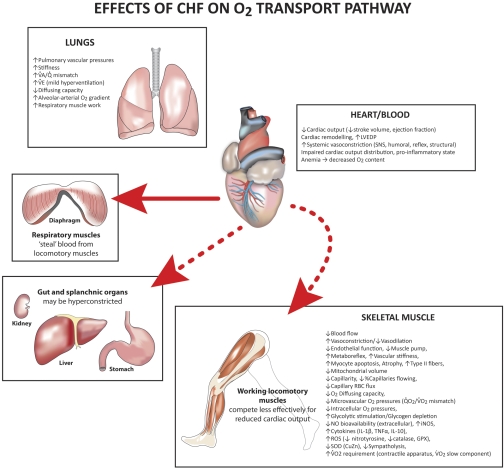
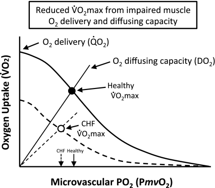
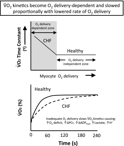
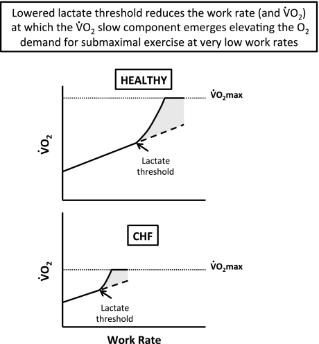
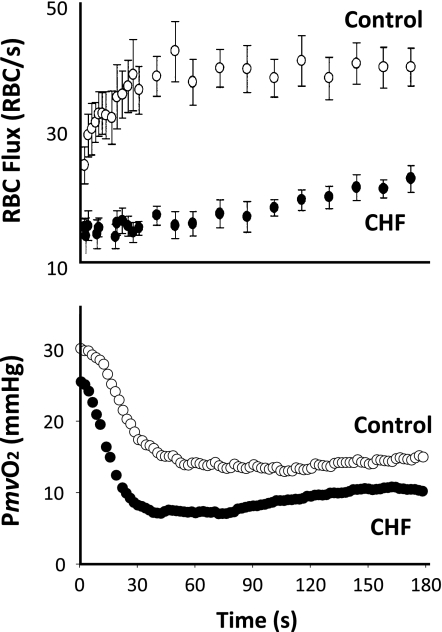
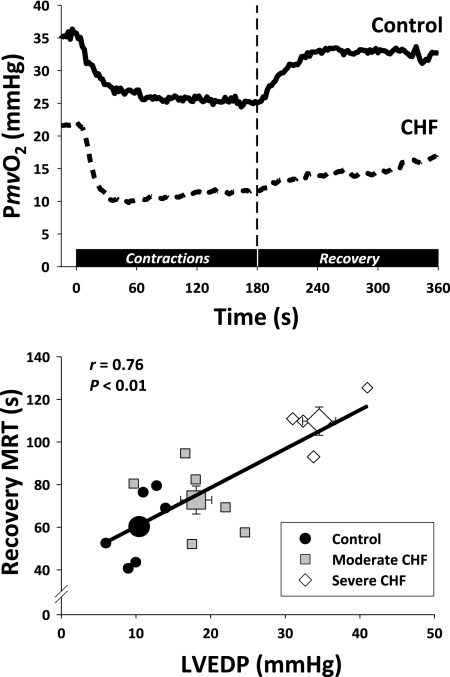
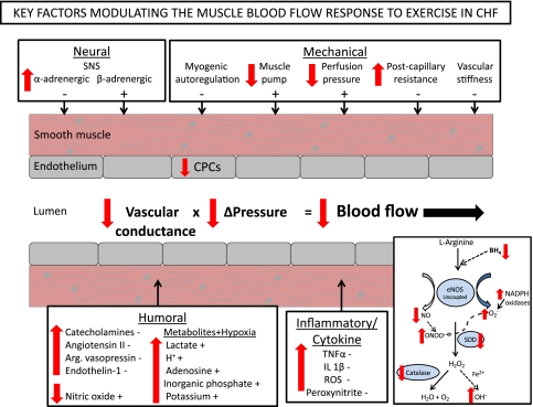
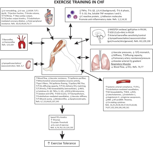
The defining characteristic of chronic heart failure (CHF) is an exercise intolerance that is inextricably linked to structural and functional aberrations in the O(2) transport pathway. CHF reduces muscle O(2) supply while simultaneously increasing O(2) demands. CHF severity varies from moderate to severe and is assessed commonly in terms of the maximum O(2) uptake, which relates closely to patient morbidity and mortality in CHF and forms the basis for Weber and colleagues' (167) classifications of heart failure, speed of the O(2) uptake kinetics following exercise onset and during recovery, and the capacity to perform submaximal exercise. As the heart fails, cardiovascular regulation shifts from controlling cardiac output as a means for supplying the oxidative energetic needs of exercising skeletal muscle and other organs to preventing catastrophic swings in blood pressure. This shift is mediated by a complex array of events that include altered reflex and humoral control of the circulation, required to prevent the skeletal muscle "sleeping giant" from outstripping the pathologically limited cardiac output and secondarily impacts lung (and respiratory muscle), vascular, and locomotory muscle function. Recently, interest has also focused on the dysregulation of inflammatory mediators including tumor necrosis factor-α and interleukin-1β as well as reactive oxygen species as mediators of systemic and muscle dysfunction. This brief review focuses on skeletal muscle to address the mechanistic bases for the reduced maximum O(2) uptake, slowed O(2) uptake kinetics, and exercise intolerance in CHF. Experimental evidence in humans and animal models of CHF unveils the microvascular cause(s) and consequences of the O(2) supply (decreased)/O(2) demand (increased) imbalance emblematic of CHF. Therapeutic strategies to improve muscle microvascular and oxidative function (e.g., exercise training and anti-inflammatory, antioxidant strategies, in particular) and hence patient exercise tolerance and quality of life are presented within their appropriate context of the O(2) transport pathway.


Figures

Fig. 1.
Fig. 1.
Predations of chronic heart failure (CHF) on the O2 transport pathway. Although a dysfunctional heart and impaired ability to generate cardiac output are the core events, CHF is a multiorgan disease affecting all steps in the O2 transport pathway. CHF-induced lung dysfunction redistributes blood flow (Q̇) to the respiratory muscles via locomotory muscle vasoconstriction; there may be systemic anemia, systemic vasoconstriction, and elevated left ventricular (LV) end-diastolic pressures (LVEDPs) as well as a plethora of structural and functional adaptations (increased vasoconstriction, impaired vasodilation, and muscle pump) that compromise skeletal muscle perfusional and diffusional O2 transport. V̇A, alveolar ventilation; V̇E, expired ventilation; SNS, sympathetic nervous system; Q̇o2, whole body oxygen delivery (cardiac output × arterial O2 content); V̇o2, oxygen uptake; NO, nitric oxide; iNOS, inducible NO synthase; ROS, reactive oxygen species; SOD, superoxide dismutase; GPX, glutathione peroxidase. See text for more details.
Fig. 2.
Fig. 2.
Facets of the exercise response in CHF: maximum V̇o2 (V̇o2 max). Schematic illustrating how the perfusive [curved lines, Fick principle, V̇o2 = muscle blood flow (Q̇m) × (arterial-venous O2 content)] and diffusive O2 [straight lines from origin, Fick's law, V̇o2 = muscle O2 capacity diffusing (Do2m) × (microvascular Po2 (PmvO2) − intracellular Po2)] transport conflate to yield the V̇o2 max during large muscle mass exercise (e.g., cycling). Note that, in CHF (dashed lines), V̇o2 max is reduced by both impaired perfusive and diffusive O2 transport and that PmvO2 may either be the same or lower (arrows on abscissa) than found in health even in the presence of marked diffusional derangements. Mechanisms responsible for these perfusive and diffusive O2 transport derangements include reduced bulk blood flow and O2 delivery, impaired blood flow distribution, reduced capillarity and percentage of capillaries supporting red blood cell (RBC) flux, lowered functional capillary hematocrit (no. of RBCs adjacent to contracting myocytes in flowing capillaries), and impaired mitochondrial function. See text for additional details.
Fig. 3.
Fig. 3.
Facets of the exercise response in CHF: V̇o2 kinetics. CHF slows V̇o2 kinetics (increased time constant, τ) in response to moderate (as shown), heavy, and severe intensity exercise, in part, by lowering muscle perfusive and diffusive O2 transport such that O2 delivery becomes limiting (top, see gray O2 delivery-dependent zone). Note that these slowed V̇o2 kinetics will mandate a greater O2 deficit leading to greater intracellular perturbations that accelerate glycogen depletion and sow the seeds for exercise intolerance. Mechanisms responsible for slowed V̇o2 kinetics in CHF include slowed/absent arteriolar vasodilation, impaired muscle pump (venous congestion), slowed capillary hemodynamics, lowered PmvO2, impaired mitochondrial function, and greater intracellular perturbation (as detailed in bottom). PCr, phosphocreatine; ADPfree, free adenosine diphosphate. See text for additional details.
Fig. 4.
Fig. 4.
Facets of the exercise response in CHF: lactate threshold (Tlac). These curves are constructed from the end-exercise V̇o2 obtained in a series of independent constant-work rate exercise tests performed in a healthy individual (top) and a patient with CHF (bottom). Note the far lower work rate for the Tlac in CHF and that the V̇o2 slow component (gray areas) becomes evident only above Tlac. One consequence of this behavior is that the patient with CHF experiences an additional O2 demand at very low work rates that may drive V̇o2 to V̇o2 max and herald imminent exhaustion. Mechanisms responsible for the lowered Tlac and presence of V̇o2 slow component at very low work rates include decreased bulk blood flow and O2 delivery, reduced capillarity, impaired capillary hemodynamics, lowered PmvO2, and mitochondrial dysfunction, particularly in slow twitch highly oxidative (type I) fibers. See text for additional details.
Fig. 5.
Fig. 5.
Top: CHF (moderate severity, LVEDP ∼10 mmHg) abolishes the rapid increase in spinotrapezius capillary RBC flux found in the healthy control muscle following onset of 1-Hz contractions (time 0 s, Ref. 136). Bottom: PmvO2 profile in the same spinotrapezius preparation. Note that in CHF PmvO2 is lower than for the healthy muscle, and there is a transient dip below the steady state (both indicative of a Q̇o2m-to-V̇o2 mismatch). From the data of Copp et al. (33), with kind permission.
Fig. 6.
Fig. 6.
Top: PmvO2 profiles for 180 s of 1-Hz contractions and 180 s of recovery for spinotrapezius muscles of healthy control and CHF rats. Note that the speed of the on-transient fall (τ) may not be substantially different but that the PmvO2 is lower at rest and throughout contractions and recovery in CHF. There is also a pronounced transient dip below the subsequent steady-state value (i.e., undershoot) for the CHF muscle. It is also striking that the recovery kinetics of the CHF muscle are markedly slowed by comparison to the on response and that of the healthy control muscle. Bottom: spinotrapezius PmvO2 recovery kinetics [mean response time (MRT), time delay + τ] was progressively slowed in CHF rats with higher LVEDPs. From Copp et al. (33), with kind permission.
Fig. 7.
Fig. 7.
Muscle blood flow during exercise in CHF is constrained by a plethora of structural, mechanical, and functional impediments that act to slow the kinetics of blood flow increase, reduce the magnitude of the exercise hyperemia, and perturb the matching between O2 delivery and V̇o2. ΔPressure, pressure differential along vessel; CPCs, circulating endothelial progenitor cells. Sign − or + indicates action to decrease or increase blood flow; red arrow gives CHF effect. Bottom, right, inset: effects of CHF on endothelial NO synthase (eNOS)-derived NO. BH4, tetrahydrobiopterin; ONOO, peroxynitrite; O2·−, superoxide; SOD, superoxide dismutase; H2O2, hydrogen peroxide; OH·−, hydroxyl radical; TNF-α, tumor necrosis factor-α; IL-1β, interleukin-1β; Arg, arginine.
Fig. 8.
Fig. 8.
Endurance exercise training opposes many of the dysfunctional elements of CHF and facilitates improved skeletal Q̇m, pulmonary gas exchange (V̇o2), and exercise tolerance. Note that the scope of these exercise training adaptations presents a substantial challenge to current and future pharmacotherapeutic approaches to treating patients with CHF (150). References cited for each organ/system are emblematic rather than comprehensive. Note that inspiratory muscle training also increases locomotory Q̇m, V̇o2 max, and exercise tolerance (27, 36, 113). dP/dt, first derivative of LV pressure; HRmax, maximum heart rate, RVLM, rostral ventrolateral medulla; NT-BNP, amino-terminal pro-brain natriuretic peptide. See text for more details.

Similar articles

Cited by

References

    1. Adamopoulos S, Parissis J, Karatzas D, Kroupis C, Georgiadis M, Karavolias G, Paraskevaidis J, Koniavitou K, Coats AJ, Kremastinos DT. Physical training modulates proinflammatory cytokines and the soluble Fas/soluble Fas ligand system in patients with chronic heart failure. J Am Coll Cardiol 39: 653–663, 2002 - PubMed
    1. Adamopoulos S, Parissis J, Kroupis C, Georgiadis M, Karatzas D, Karavolias G, Koniavitou K, Coats AJ, Kremastinos DT. Physical training reduces peripheral markers of inflammation in patients with chronic heart failure. Eur Heart J 22: 791–797, 2001 - PubMed
    1. Adams V, Jiang H, Yu J, Möbius-Winkler S, Fiehn E, Linke A, Weigl C, Schuler G, Hambrecht R. Apoptosis in skeletal myocytes of patients with chronic heart failure is associated with exercise intolerance. J Am Coll Cardiol 33: 959–965, 1999 - PubMed
    1. Adams V, Nehrhoff B, Späte U, Linke A, Schulze PC, Baur A, Gielen S, Hambrecht R, Schuler G. Induction of iNOS expression in skeletal muscle by IL-1beta and NFkappaB activation: an in vitro and in vivo study. Cardiovasc Res 54: 95–104, 2002 - PubMed
    1. Agostoni PG, Bussotti M, Palermo P, Guazzi M. Does lung diffusion impairment affect exercise capacity in patients with heart failure? Heart 88: 453–459, 2002 - PMC - PubMed

Publication types