. 2011 Nov 20;17(12):1646-51.
doi: 10.1038/nm.2580.

Functionally recurrent rearrangements of the MAST kinase and Notch gene families in breast cancer

Affiliations

Functionally recurrent rearrangements of the MAST kinase and Notch gene families in breast cancer

Dan R Robinson et al. Nat Med. .

Abstract

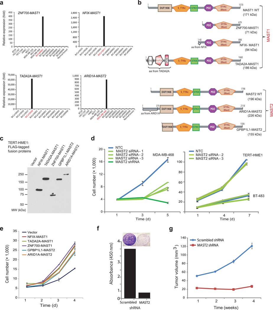
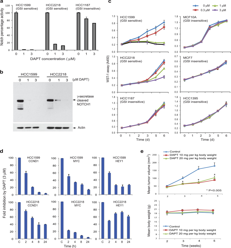
Breast cancer is a heterogeneous disease that has a wide range of molecular aberrations and clinical outcomes. Here we used paired-end transcriptome sequencing to explore the landscape of gene fusions in a panel of breast cancer cell lines and tissues. We observed that individual breast cancers have a variety of expressed gene fusions. We identified two classes of recurrent gene rearrangements involving genes encoding microtubule-associated serine-threonine kinase (MAST) and members of the Notch family. Both MAST and Notch-family gene fusions have substantial phenotypic effects in breast epithelial cells. Breast cancer cell lines harboring Notch gene rearrangements are uniquely sensitive to inhibition of Notch signaling, and overexpression of MAST1 or MAST2 gene fusions has a proliferative effect both in vitro and in vivo. These findings show that recurrent gene rearrangements have key roles in subsets of carcinomas and suggest that transcriptome sequencing could identify individuals with rare, targetable gene fusions.


Figures

Figure 1
Figure 1. Discovery of the MAST kinase and Notch gene fusions in breast cancer identified by paired-end transcriptome sequencing
(a) MAST family gene fusions. (b) Notch family gene fusions. Fusion junctions with respective exon numbers (and nucleotide positions) comprising the chimeric transcripts are presented. Bar plots of top ranked gene fusions by number of paired-end reads supporting each nominated fusion in the index samples are shown on the right, with MAST or Notch fusion genes in red.
Figure 2
Figure 2. Characterization of MAST fusion genes
(a) Expression of ZNF700-MAST1 gene fusion in breast cancer tissue BrCa00001, NFIX-MAST1 in BrCa10017, TADA2AMAST1 fusion in BrCa10038, and ARID1A-MAST2 fusion in MDA-MB-468 was validated by real-time RT-PCR. (b) Schematic representation of functional domains retained in the putative chimeric proteins involving MAST1 and MAST2. (c) Stable expression of MAST fusion proteins in TERT-HME1 cells. The expression of the chimeric proteins was detected by anti-FLAG antibody. (d) MAST2 knockdown reduces proliferation of MDA-MB-468 cells (left) but not fusion-negative TERT-HME1 and BT-483 cells (right). (e) Overexpression of MAST chimera induces cell proliferation in TERT-HME1 cells. (f) Colony formation assay with MDA-MB-468 cells treated with MAST2-specific shRNA or control scrambled-shRNA. The inset shows crystal violet staining of cells treated with either scrambled or MAST2 shRNA. (g) Reduced tumor growth by MAST2 knockdown in a mouse xenograft model.
Figure 3
Figure 3. Identification and characterization of Notch gene aberrations in breast carcinomas
(a) Detection of novel Notch transcripts by quantitative RT-PCR. (b) Schematic presentation of the predicted protein structures of the aberrant Notch genes. (c) Notch reporter activities are elevated in Notch fusion index lines. Fold activation of Notch pathway was calculated using HCC202 as the reference. All the data was normalized to Renilla luciferase activity. (d) Western blot analysis of NOTCH1-NICD expression, detected with an antibody specifically recognizing the active NOTCH1-NICD protein after γ-secretase cleavage. FL, full length NOTCH1; TM, transmembrane NOTCH1. (e) Activation of Notch signaling pathway in 293T cells by transient Notch expression. (f) Notch fusion alleles induce morphological change when expressed in benign TERT-HME1 cells. Bright-field images of control vector, Notch fusion allele expressing TERT-HME1 cells. Notch fusion positive breast cancer lines are shown for comparison. (g) Activation of Notch signaling pathway in TERT-HME1 cells stably expressing Notch fusions. The expression levels of three Notch target genes were measured by quantitative RT-PCR.
Figure 4
Figure 4. γ-secretase inhibitor DAPT blocks Notch-dependent cell proliferation
(a) Inhibition of the Notch signaling pathway by DAPT. Breast cancer cells were co-infected with a Notch-reporter construct Lenti-RBPJ-firefly luciferase and the internal control Lenti-Renilla luciferase and treated with DAPT. Twenty-four hours after DAPT treatment, luciferase activities were measured. (b) Reduction of NICD production after DAPT treatment, detected with an antibody specific to the active NOTCH1-NICD after γ-secretase cleavage. (c) Inhibition of cell proliferation by DAPT. HCC1599 and HCC2218 express a form of NOTCH1 which requires γ-secretase-dependent cleavage for activity. HCC1187 cells express a NOTCH2-fusion mutant that is independent of γ–secretase processing. MCF10A, MCF7, and HCC1395 express wild type NOTCH1 and NOTCH2. GSI: γ-secretase inhibitor. (d) Diminished expression of Notch target genes following DAPT treatment, measured by quantitative RT-PCR. (e) Inhibition of tumor growth by DAPT in a mouse xenograft model. Mice xenografted with HCC1599 cells were treated with DAPT after tumors were formed, and the tumor size was monitored.

References

    1. Delattre O, et al. Gene fusion with an ETS DNA-binding domain caused by chromosome translocation in human tumours. Nature. 1992;359:162–165. - PubMed
    1. Nowell PC, Hungerford DA. Chromosome studies on normal and leukemic human leukocytes. J Natl Cancer Inst. 1960;25:85–109. - PubMed
    1. Rowley JD. The critical role of chromosome translocations in human leukemias. Annu Rev Genet. 1998;32:495–519. - PubMed
    1. Kumar-Sinha C, Tomlins SA, Chinnaiyan AM. Recurrent gene fusions in prostate cancer. Nat Rev Cancer. 2008;8:497–511. - PMC - PubMed
    1. Tomlins SA, et al. Recurrent fusion of TMPRSS2 and ETS transcription factor genes in prostate cancer. Science. 2005;310:644–648. - PubMed

Publication types

MeSH terms