Comparative Study
. 2011 Nov;67(Pt 11):907-14.
doi: 10.1107/S0907444911035943. Epub 2011 Oct 19.

High-resolution structure of a retroviral protease folded as a monomer

Affiliations
Comparative Study

High-resolution structure of a retroviral protease folded as a monomer

Miroslaw Gilski et al. Acta Crystallogr D Biol Crystallogr. 2011 Nov.

Abstract

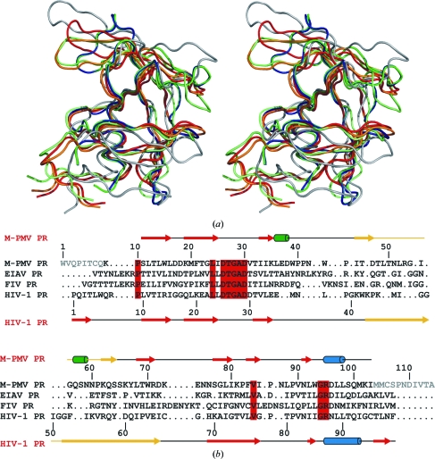
Mason-Pfizer monkey virus (M-PMV), a D-type retrovirus assembling in the cytoplasm, causes simian acquired immunodeficiency syndrome (SAIDS) in rhesus monkeys. Its pepsin-like aspartic protease (retropepsin) is an integral part of the expressed retroviral polyproteins. As in all retroviral life cycles, release and dimerization of the protease (PR) is strictly required for polyprotein processing and virion maturation. Biophysical and NMR studies have indicated that in the absence of substrates or inhibitors M-PMV PR should fold into a stable monomer, but the crystal structure of this protein could not be solved by molecular replacement despite countless attempts. Ultimately, a solution was obtained in mr-rosetta using a model constructed by players of the online protein-folding game Foldit. The structure indeed shows a monomeric protein, with the N- and C-termini completely disordered. On the other hand, the flap loop, which normally gates access to the active site of homodimeric retropepsins, is clearly traceable in the electron density. The flap has an unusual curled shape and a different orientation from both the open and closed states known from dimeric retropepsins. The overall fold of the protein follows the retropepsin canon, but the C(α) deviations are large and the active-site 'DTG' loop (here NTG) deviates up to 2.7 Å from the standard conformation. This structure of a monomeric retropepsin determined at high resolution (1.6 Å) provides important extra information for the design of dimerization inhibitors that might be developed as drugs for the treatment of retroviral infections, including AIDS.


Figures

Figure 1
Figure 1
Stereoview of the main-chain trace of the flap loop plus flanking residues (Trp43–Tyr67). This trace of the flap of molecule A is shown in 2F oF c electron density contoured at 1.0σ. Side-chain atoms have been omitted for clarity.
Figure 2
Figure 2
Alignment of retroviral proteases. (a) Stereoview of the superposition of the Cα traces of protomers of retroviral proteases: green and blue, M-PMV (A and B); red, HIV-1, apo form (PDB entry 3hvp); orange, HIV-1, inhibitor complex (PDB entry 4hvp); lime, EIAV (PDB entry 2fmb); grey, M-PMV, NMR model (PDB entry 1nso), energy-minimized (in water). (b) Structure-based sequence alignment of the M-PMV, EIAV (PDB entry 2fmb; lowest core Cα r.m.s.d.; Table 2 ▶), FIV (PDB entry 4fiv; highest level of sequence identity – 26.6%) and HIV-1 (PDB entry 3hvp) proteases. Residue numbers and secondary-structure elements (arrows, β-strands; blue, α-helices; green, 310-helices; yellow, flap loops) are marked for the M-­PMV and HIV-1 proteases. Residues that are identical in all four sequences are shown on a red background. Disordered residues missing from the M-PMV PR structure are shown in grey.
Figure 3
Figure 3
Stereoview of overlay of the active-site (D/N)TG loops of HIV-1 PR (PDB entry 3hvp, grey) and M-PMV PR (green) based on Cα superposition of the entire molecules. The M-PMV PR structure is shown as 2F oF c electron density contoured at 1.3σ.
Figure 4
Figure 4
Electrostatic potential surface of retroviral protease protomers. The M-PMV PR monomer (a) is shown in the same orientation and on the same scale as the HIV-1 PR protomer (b) extracted from the dimeric molecule (PDB entry 3hvp). The complete HIV-1 PR dimer is generated by the action of a vertical dyad, which creates a second copy facing the first molecule on the right. In this view, the N- and C-­termini (missing in M-PMV PR) are at the bottom and the flap loops are at the top. The active-site cavity is marked by the Asn26/Asp25 residue (ball-and-stick representation). In M-PMV PR the cavity is completely covered by the curled flap. The area of positive potential on this M-PMV PR surface is influenced by the D26N substitution, but it is of note that this mutation does not influence the tendency of the protein to fold as a monomer. The electrostatic potential (negative, red; positive, blue) was calculated in APBS (Baker et al., 2001 ▶).
Figure 5
Figure 5
Stereoview of superposition of the Cα atoms of the HIV-1 PR protomer (PDB entry 3hvp, red) on monomer A of M-PMV PR in the crystal structure (green). This superposition illustrates the relation of the HIV-1 PR dimer (cartoon) to the neighbouring copies of M-PMV PR monomer B in the crystal (blue and magenta). Bottom panel, view down the twofold axis of the HIV-1 PR dimer; top panel, a perpendicular view with the twofold axis vertical.

Similar articles

Cited by

References

    1. Adams, P. D. et al. (2010). Acta Cryst. D66, 213–221. - PubMed
    1. Baker, N. A., Sept, D., Joseph, S., Holst, M. J. & McCammon, J. A. (2001). Proc. Natl Acad. Sci. USA, 98, 10037–10041. - PMC - PubMed
    1. Bauerová-Zábranská, H., Stokrová, J., Strísovsky, K., Hunter, E., Ruml, T. & Pichová, I. (2005). J. Biol. Chem. 280, 42106–42112. - PubMed
    1. Cohen, G. H. (1997). J. Appl. Cryst. 30, 1160–1161.
    1. Cooper, S., Khatib, F., Treuille, A., Barbero, J., Lee, J., Beenen, M., Leaver-Fay, A., Baker, D. & Popović, Z. (2010). Nature (London), 466, 756–760. - PMC - PubMed

Publication types

MeSH terms

Substances

Associated data