. 2011 Nov;67(Pt 11):920-8.
doi: 10.1107/S0907444911036353. Epub 2011 Oct 19.

Crystallographic analysis of human hemoglobin elucidates the structural basis of the potent and dual antisickling activity of pyridyl derivatives of vanillin

Affiliations

Crystallographic analysis of human hemoglobin elucidates the structural basis of the potent and dual antisickling activity of pyridyl derivatives of vanillin

Osheiza Abdulmalik et al. Acta Crystallogr D Biol Crystallogr. 2011 Nov.

Erratum in

  • Acta Crystallogr D Biol Crystallogr. 2011 Dec;67(Pt 12):1076

Abstract

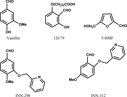
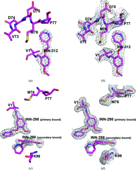
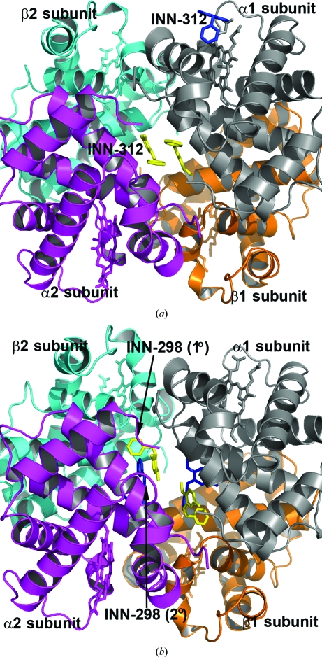
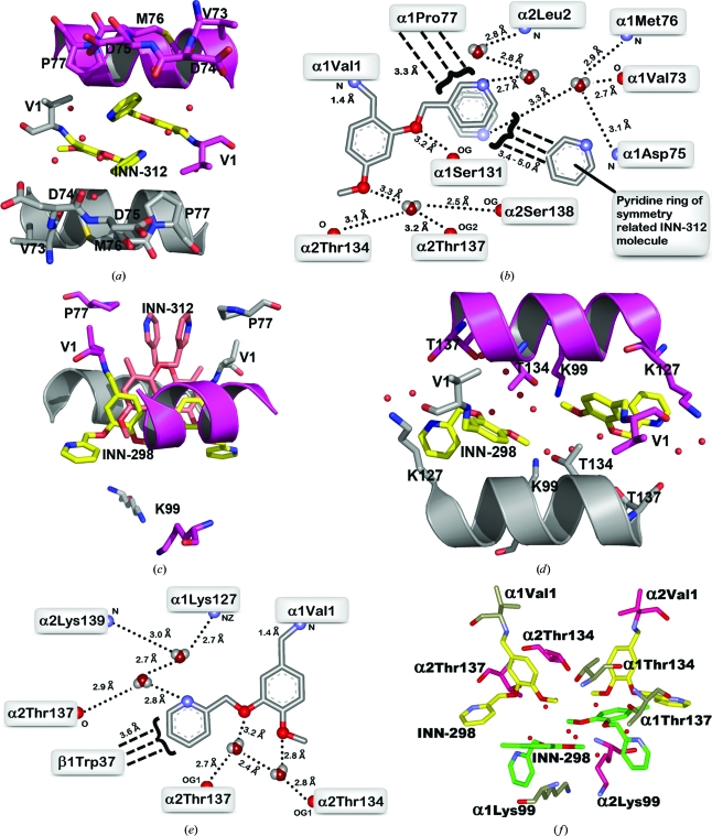
Vanillin has previously been studied clinically as an antisickling agent to treat sickle-cell disease. In vitro investigations with pyridyl derivatives of vanillin, including INN-312 and INN-298, showed as much as a 90-fold increase in antisickling activity compared with vanillin. The compounds preferentially bind to and modify sickle hemoglobin (Hb S) to increase the affinity of Hb for oxygen. INN-312 also led to a considerable increase in the solubility of deoxygenated Hb S under completely deoxygenated conditions. Crystallographic studies of normal human Hb with INN-312 and INN-298 showed that the compounds form Schiff-base adducts with the N-terminus of the α-subunits to constrain the liganded (or relaxed-state) Hb conformation relative to the unliganded (or tense-state) Hb conformation. Interestingly, while INN-298 binds and directs its meta-positioned pyridine-methoxy moiety (relative to the aldehyde moiety) further down the central water cavity of the protein, that of INN-312, which is ortho to the aldehyde, extends towards the surface of the protein. These studies suggest that these compounds may act to prevent sickling of SS cells by increasing the fraction of the soluble high-affinity Hb S and/or by stereospecific inhibition of deoxygenated Hb S polymerization.


Figures

Figure 1
Figure 1
Structures of vanillin, 12C79, 5-HMF and INN compounds.
Figure 2
Figure 2
An initial difference electron-density map (with coefficients F oF c shown at the 2.5σ level) of the R2-state INN-312 complex structure before the bound INN-312 molecules at the α-cleft were added to the model. Only one of the two bound molecules is shown. (b) A final electron-density map (with coefficients 2F oF c shown at the 0.8σ level) of the R2-state INN-312 complex structure. (c) An initial difference electron-density map (with coefficients F oF c shown at the 2.2σ level) of the R2-state INN-298 complex structure calculated before the primary and secondary bound INN-298 molecules at the α-cleft were added to the model. (d) A final electron-density map (with coefficients 2F oF c shown at the 0.8σ level) of the R2-state INN-312 complex structure. All maps are superimposed with the final refined models.
Figure 3
Figure 3
Crystal structures of R2-state Hb in complex with two molecules of INN-312 bound at the α-cleft (yellow sticks) and a third molecule of INN-312 (blue sticks) bound at a surface cavity near the α1 heme binding pocket. Hb subunits are shown as ribbons (α1 subunit in ash, α2 subunit in magenta, β1 subunit in orange and β2 subunit in cyan) and hemes are shown as sticks. (b) Primary bound (yellow sticks) and secondary bound (blue sticks) INN-298 molecules at the α-cleft in the R2-state structure.
Figure 4
Figure 4
A pair of INN-312 molecules (yellow sticks) bound at the α-cleft of R2-state Hb making Schiff-base interactions with αVal1 N. Hb subunits are shown as sticks or ribbons (α1 subunit in ash, α2 subunit in magenta). Water molecules are shown as red spheres. For brevity, not all residues that make contact with the compound are shown (interacting residues are shown in Fig. 4 ▶ b). (b) Two-dimensional contacts between one of the bound INN-312 molecules and the protein at the α-cleft. Small-dashed lines indicate hydrogen-bond interactions and broad-dashed lines indicate hydrophobic contacts. Note the rotation of the pyridine ring with respect to the aryl–C bond, which allows the 3-pyridine N atom to make alternate water-mediated hydrogen-bond contacts with the protein. (c) Superimposition of the primary bound INN-298 (yellow) and INN-312 (brown) molecules in R2-state Hb. The protein residues are from the INN-298 structure. (d) A pair of INN-298 molecules (yellow sticks) bound at the α-cleft of the R2-state Hb making Schiff-base interactions with αVal1 N. There is a second pair of symmetry-related INN-298 molecules bound further down the central water cavity (see Fig. 4 ▶ f). (e) Two-dimensional contacts between one of the primary bound INN-298 molecules and the protein at the α-cleft. (f) Primary (yellow sticks) and secondary (green sticks) bound INN-298 molecules.

References

    1. Abdulmalik, O., Safo, M. K., Chen, Q., Yang, J., Brugnara, C., Ohene-Frempong, K., Abraham, D. J. & Asakura, T. (2005). Br. J. Haematol. 128, 552–561. - PubMed
    1. Abraham, D. J., Mehanna, A. S., Wireko, F. C., Whitney, J., Thomas, R. P. & Orringer, E. P. (1991). Blood, 77, 1334–1341. - PubMed
    1. Abraham, D. J., Safo, M. K., Boyiri, T., Danso-Danquah, R. E., Kister, J. & Poyart, C. (1995). Biochemistry, 34, 15006–15020. - PubMed
    1. Beddell, C. R., Goodford, P. J., Kneen, G., White, R. D., Wilkinson, S. & Wootton, R. (1984). Br. J. Pharmacol. 82, 397–407. - PMC - PubMed
    1. Boyiri, T., Safo, M. K., Danso-Danquah, R. E., Kister, J., Poyart, C. & Abraham, D. J. (1995). Biochemistry, 34, 15021–15036. - PubMed

Publication types

MeSH terms