. 2012 Feb;194(3):567-75.
doi: 10.1128/JB.05553-11. Epub 2011 Nov 18.

Iron storage proteins are essential for the survival and pathogenesis of Mycobacterium tuberculosis in THP-1 macrophages and the guinea pig model of infection

Affiliations

Iron storage proteins are essential for the survival and pathogenesis of Mycobacterium tuberculosis in THP-1 macrophages and the guinea pig model of infection

P Vineel Reddy et al. J Bacteriol. 2012 Feb.

Abstract

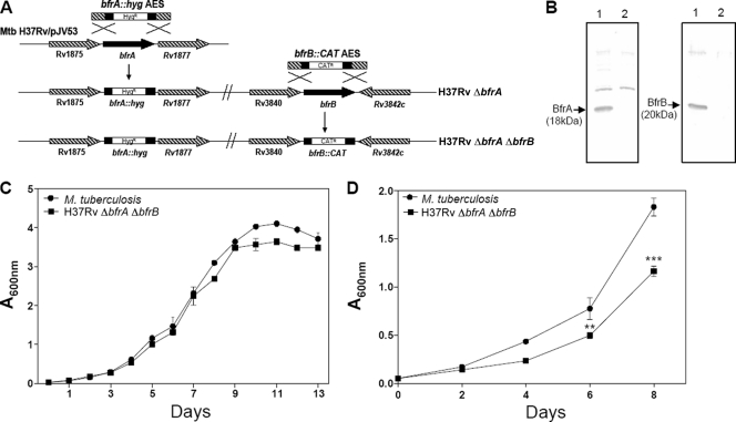
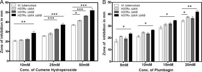
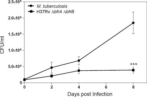
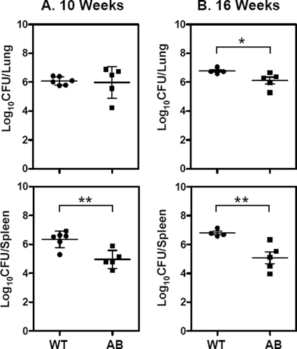
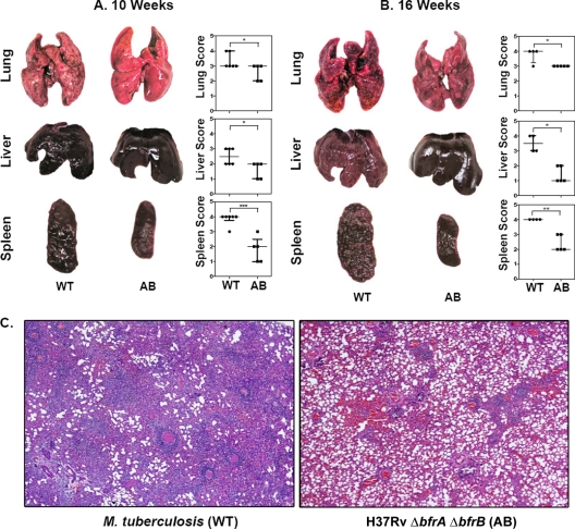
Iron is one of the crucial elements required for the growth of Mycobacterium tuberculosis. However, excess free iron becomes toxic for the cells because it catalyzes the production of reactive oxygen radicals, leading to oxidative damage. Hence, it is essential for the pathogen to have the ability to store intracellular iron in an iron-rich environment and utilize it under iron depletion. M. tuberculosis has two iron storage proteins, namely BfrA (Rv1876; a bacterioferritin) and BfrB (Rv3841; a ferritin-like protein). However, the demonstration of biological significance requires the disruption of relevant genes and the evaluation of the resulting mutant for its ability to survive in the host and cause disease. In this study, we have disrupted bfrA and bfrB of M. tuberculosis and demonstrated that these genes are crucial for the storage and supply of iron for the growth of bacteria and to withstand oxidative stress in vitro. In addition, the bfrA bfrB double mutant (H37Rv ΔbfrA ΔbfrB) exhibited a marked reduction in its ability to survive inside human macrophages. Guinea pigs infected with H37Rv ΔbfrA ΔbfrB exhibited a marked diminution in the dissemination of the bacilli to spleen compared to that of the parental strain. Moreover, guinea pigs infected with H37Rv ΔbfrA ΔbfrB exhibited significantly reduced pathological damage in spleen and lungs compared to that of animals infected with the parental strain. Our study clearly demonstrates the importance of these iron storage proteins in the survival and pathogenesis of M. tuberculosis in the host and establishes them as attractive targets for the development of new inhibitors against mycobacterial infections.


Figures

Fig 1
Fig 1
Characterization of H37Rv ΔbfrA ΔbfrB. (A) Disruption of bfrA and bfrB genes of M. tuberculosis by using the recombineering method. The diagram represents homologous recombination between bfrA::hyg AES and the bfrA gene in M. tuberculosis to generate H37Rv ΔbfrA, followed by recombination between bfrB::CAT AES and the bfrB gene in H37Rv ΔbfrA to generate H37Rv ΔbfrA ΔbfrB. (B) Confirmation of disruption of bfrA and bfrB deletion in M. tuberculosis by immunoblotting. Five μg of cell-free protein extract of M. tuberculosis (lane 1) and H37Rv ΔbfrA ΔbfrB (lane 2) was loaded onto a 12% polyacrylamide gel and subjected to electrophoresis. BfrA and BfrB were detected by immunoblot analysis using anti-BfrA or anti-BfrB polyclonal antiserum. BfrA and BfrB proteins migrated as protein bands corresponding to a molecular mass of 18 and 20 kDa, respectively (lane 1). The disruption of bfrA and bfrB in H37Rv ΔbfrA ΔbfrB was confirmed by the lack of expression of either protein (lane 2). (C) Growth kinetics of M. tuberculosis and H37Rv ΔbfrA ΔbfrB in MB7H9 medium. Cultures were inoculated in duplicate with a starting absorbance (A600) of 0.025, and the growth was monitored for 13 days. (D) Growth curve of M. tuberculosis H37Rv and H37Rv ΔbfrA ΔbfrB in iron-depleted medium. M. tuberculosis H37Rv and H37Rv ΔbfrA ΔbfrB were grown in MB7H9 medium supplemented with 0.05% Tween 80, 10× ADC, and 100 μM 2′2′ dipyridyl. The growth of the strains was monitored by measuring the A600 for 8 days. There was a significant difference in the growth of H37Rv ΔbfrA ΔbfrB compared to that of M. tuberculosis H37Rv under iron-deprived conditions. The values of absorbance are represented as the means (± standard errors) of three independent samples, and the experiment was repeated three times. **, P < 0.01; ***, P < 0.001 (two-way ANOVA).
Fig 2
Fig 2
Influence of disruption of bfrA and bfrB on the ability of M. tuberculosis to withstand oxidative stress. Shown is the range of the inhibition zones observed in the presence of various concentrations of cumene hydroperoxide (A) and plumbagin (B). H37Rv ΔbfrA exhibited a significant sensitivity toward both cumene hydroperoxide and plumbagin, although only at higher concentrations, whereas H37Rv ΔbfrB exhibited a significant sensitivity toward cumene hydroperoxide. H37Rv ΔbfrA ΔbfrB exhibited a significantly higher sensitivity to oxidative stress induced by both agents compared to that of the parental strain. The values of zones of inhibition are represented as the means (± standard errors) of three independent samples, and the experiment was repeated three times. *, P < 0.05; **, P < 0.01; ***, P < 0.001 (two-way ANOVA).
Fig 3
Fig 3
Influence of deletion of bfrA and bfrB genes on the growth of M. tuberculosis in THP-1 cells. THP-1 cells were infected with M. tuberculosis or the H37Rv ΔbfrA ΔbfrB mutant at an MOI of 1:5 (bacterium to macrophage), and the number of intracellular viable bacteria was determined for 8 days. Cells were lysed, and appropriate dilutions of mycobacteria were plated onto MB7H11 agar to determine the CFU. The H37Rv ΔbfrA ΔbfrB mutant exhibited a marked attenuation in its growth compared to that of M. tuberculosis H37Rv at 8 days postinfection. The values are represented as the means (± standard errors) of three independent infections, and the experiment was repeated three times. ***, P < 0.001 (two-way ANOVA).
Fig 4
Fig 4
Influence of disruption of bfrA and bfrB genes of M. tuberculosis on the growth of the pathogen in guinea pigs. The figure depicts the bacillary load in the lungs and spleen of guinea pigs (n = 6) infected with M. tuberculosis (WT) and the H37Rv ΔbfrA ΔbfrB mutant (AB) at 10 (A) and 16 (B) weeks postinfection. Guinea pigs infected with H37Rv ΔbfrA ΔbfrB (AB) exhibited a significantly reduced bacillary load in spleen compared to animals infected with M. tuberculosis (WT). Each data point represents the log10 CFU value for an individual animal, and the bars depict means (± standard errors) for each group. Missing data points represent the animals that succumbed to disease before the time of euthanasia. *, P < 0.05; **, P < 0.01 (Student's t test).
Fig 5
Fig 5
Influence of disruption of bfrA and bfrB genes of M. tuberculosis on gross pathological lesions and histopathological damage in organs of infected guinea pigs. The figure depicts representative photographs of gross pathological lesions in lung, liver, and spleen of guinea pigs (n = 6) infected with M. tuberculosis (WT) and H37Rv ΔbfrA ΔbfrB (AB) euthanized at 10 (A) and 16 (B) weeks postinfection. Guinea pigs infected with H37Rv ΔbfrAΔbfrB resulted in fewer and smaller lung, liver, and spleen lesions compared to animals infected with M. tuberculosis. Each data point represents the score of an individual animal, and the bars depict medians (± interquartile ranges) for each group. Missing data points represent the animals that succumbed to disease before the time of euthanasia. *, P < 0.05; **, P < 0.01; ***, P < 0.001 (Student's t test). (C) Influence of the disruption of bfrA and bfrB genes of M. tuberculosis on the histopathological damage to the organs of infected guinea pigs. The lung tissues were fixed in 10% buffered formalin and were embedded in paraffin. Subsequently, 5-μm-thick sections were cut and stained with hematoxylin and eosin (H&E) for histopathological examination. The figure depicts a representative photograph of the extent of pathological damage to animals (n = 6) infected with either M. tuberculosis (A) or H37Rv ΔbfrA ΔbfrB (B) at ×40 magnification at 10 weeks postinfection. H37Rv ΔbfrA ΔbfrB-infected animals showed reduced granulomatous infiltration, with only a few small and discrete granulomas, compared to that of M. tuberculosis-infected animals.
Fig 6
Fig 6
Influence of disruption of bfrA and bfrB genes of M. tuberculosis on the survival of guinea pigs postinfection. Guinea pigs infected with M. tuberculosis H37Rv/pJV53 or H37Rv ΔbfrA ΔbfrB through the aerosol route were monitored for survival (n = 6). The experiment was started with a bacillary load of 5 to 10 CFU in lungs at 1 day postinfection. Bacillary burden in lungs and spleen was monitored at 5, 10, and 16 weeks postinfection. Remaining guinea pigs (n = 6) were monitored for survival up to 21 weeks postinfection.

Similar articles

Cited by

References

    1. Abdul-Tehrani H, et al. 1999. Ferritin mutants of Escherichia coli are iron deficient and growth impaired, and fur mutants are iron deficient. J. Bacteriol. 181:1415–1428 - PMC - PubMed
    1. Almirón MA, Ugalde RA. 2010. Iron homeostasis in Brucella abortus: the role of bacterioferritin. J. Microbiol. 48:668–673 - PubMed
    1. Andrews SC. 1998. Iron storage in bacteria. Adv. Microb. Physiol. 40:281–351 - PubMed
    1. Bardarov S, et al. 2002. Specialized transduction: an efficient method for generating marked and unmarked targeted gene disruptions in Mycobacterium tuberculosis, M. bovis BCG, and M. smegmatis. Microbiology 148:3007–3017 - PubMed
    1. Boelaert JR, Vandecasteele SJ, Appelberg R, Gordeuk VR. 2007. The effect of the host's iron status on tuberculosis. J. Infect. Dis. 195:1745–1753 - PubMed

Publication types