Comparative Study
. 2012 Feb;194(3):702-7.
doi: 10.1128/JB.06184-11. Epub 2011 Nov 18.

Unveiling unusual features of formation of septal partition and constriction in mycobacteria--an ultrastructural study

Affiliations
Comparative Study

Unveiling unusual features of formation of septal partition and constriction in mycobacteria--an ultrastructural study

Srinivasan Vijay et al. J Bacteriol. 2012 Feb.

Abstract

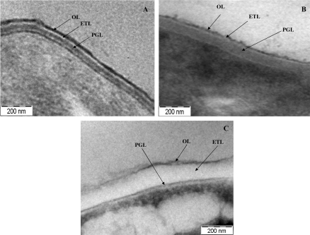
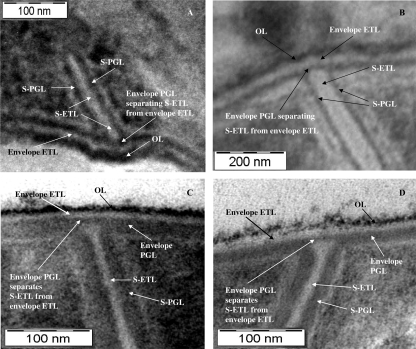
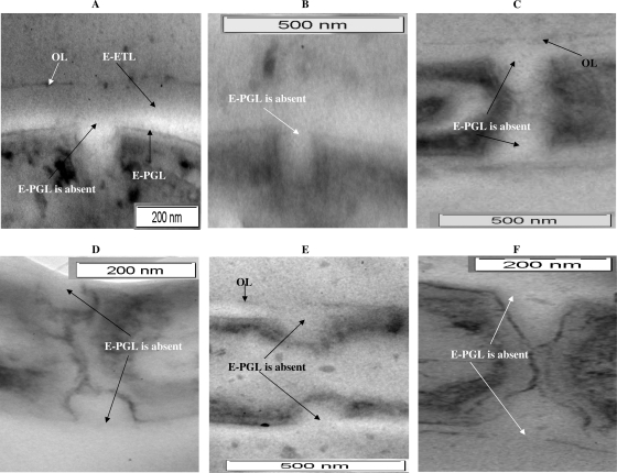
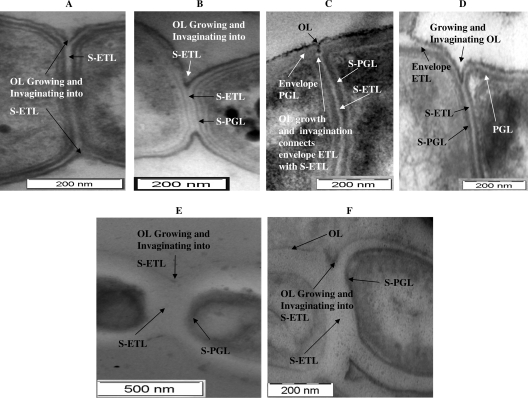
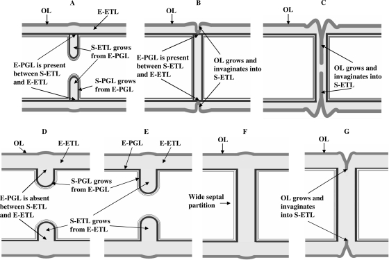
The ultrastructural functions of the electron-dense glycopeptidolipid-containing outermost layer (OL), the arabinogalactan-mycolic acid-containing electron-transparent layer (ETL), and the electron-dense peptidoglycan layer (PGL) of the mycobacterial cell wall in septal growth and constriction are not clear. Therefore, using transmission electron microscopy, we studied the participation of the three layers in septal growth and constriction in the fast-growing saprophytic species Mycobacterium smegmatis and the slow-growing pathogenic species Mycobacterium xenopi and Mycobacterium tuberculosis in order to document the processes in a comprehensive and comparative manner and to find out whether the processes are conserved across different mycobacterial species. A complete septal partition is formed first by the fresh synthesis of the septal PGL (S-PGL) and septal ETL (S-ETL) from the envelope PGL (E-PGL) in M. smegmatis and M. xenopi. The S-ETL is not continuous with the envelope ETL (E-ETL) due to the presence of the E-PGL between them. The E-PGL disappears, and the S-ETL becomes continuous with the E-ETL, when the OL begins to grow and invaginate into the S-ETL for constriction. However, in M. tuberculosis, the S-PGL and S-ETL grow from the E-PGL and E-ETL, respectively, without a separation between the E-ETL and S-ETL by the E-PGL, in contrast to the process in M. smegmatis and M. xenopi. Subsequent growth and invagination of the OL into the S-ETL of the septal partition initiates and completes septal constriction in M. tuberculosis. A model for the conserved sequential process of mycobacterial septation, in which the formation of a complete septal partition is followed by constriction, is presented. The probable physiological significance of the process is discussed. The ultrastructural features of septation and constriction in mycobacteria are unusually different from those in the well-studied organisms Escherichia coli and Bacillus subtilis.


Figures

Fig 1
Fig 1
Ultrastructures of cell envelopes of M. smegmatis (A), M. xenopi (B), and M. tuberculosis (C). OL, outer layer; ETL, electron-transparent layer; PGL, peptidoglycan layer.
Fig 2
Fig 2
Ultrastructural profile of the initial stages of formation of the septal partition in M. smegmatis (A and B) and M. xenopi (C and D). The S-PGL and S-ETL grow from the E-PGL. The S-PGL is connected to the E-PGL, but the S-ETL is separated from the E-ETL by the presence of the E-PGL. The S-ETL is flanked by the S-PGL (one layer on each side).
Fig 3
Fig 3
Images of initial stages of formation of the septal partition in M. tuberculosis cells. The S-PGL and S-ETL grow continuously with the E-PGL and E-ETL, respectively. The E-PGL is not present between the S-ETL and E-ETL. Note that in the very early stage of the initiation of septal partition, the S-PGL and S-ETL grow continuously with, and connected to, the E-PGL and E-ETL, respectively (A).
Fig 4
Fig 4
Ultrastructural features of the initiation of constriction. (A, C, and E) The OL begins to grow and invaginate into the S-ETL in M. smegmatis, M. xenopi, and M. tuberculosis, respectively. The E-PGL separates the S-ETL from the E-ETL during septation in M. smegmatis and M. xenopi. This separation disappears when the OL invaginates into the S-ETL. This makes the E-ETL continuous with the S-ETL. In contrast, no such temporary separation of the S-ETL from the E-ETL by the E-PGL seems to occur in M. tuberculosis. The S-ETL is flanked by the S-PGL (one layer on each side). (B, D, and F) Later stages of growth and invagination of the OL into the septal ETL in M. smegmatis, M. xenopi, and M. tuberculosis, respectively.
Fig 5
Fig 5
Model for formation of the septal partition and constriction in M. smegmatis, M. xenopi, and M. tuberculosis. (A to C) M. smegmatis and M. xenopi. (A) The S-ETL and S-PGL grow, connected to the E-PGL, which separates the S-ETL from the E-ETL. (B) Completion of formation of the septal partition constituted by the S-ETL and S-PGL. The continued presence of the E-PGL, separating the S-ETL from the E-ETL, is shown. The OL starts to grow and invaginate into the S-ETL to form constriction. (C) Progression of growth and invagination of the OL into the S-ETL. (D to G) M. tuberculosis. (D) The S-ETL and S-PGL grow continuously with the E-ETL and E-PGL, respectively. In contrast to the process in M. smegmatis and M. xenopi, the E-PGL is not present between the S-ETL and E-ETL. (F) Completion of formation of septal partition. (G) The OL begins to grow and invaginate into the S-ETL to form constriction.

References

    1. Barksdale L, Kim K-S. 1977. Mycobacterium. Bacteriol. Rev. 41:217–372 - PMC - PubMed
    1. Brennan PJ, Nikaido H. 1995. The envelope of mycobacteria. Annu. Rev. Biochem. 64:29–63 - PubMed
    1. Brennan PJ. 2003. Structure, function, and biogenesis of the cell wall of Mycobacterium tuberculosis. Tuberculosis 83:91–97 - PubMed
    1. Conti SF, Gettner EM. 1962. Electron microscopy of cellular division in Escherichia coli. J. Bacteriol. 83:544–550 - PMC - PubMed
    1. Crick DC, Quadri L, Brennan PJ. 2008. Biochemistry of the cell envelope of Mycobacterium tuberculosis, p 1–19 In Kaufmann SHE, Rubin E. (ed), Handbook of tuberculosis: molecular biology and biochemistry. Wiley-VCH Verlag GmbH & Co KGaA, Weinheim, Germany

Publication types

MeSH terms

LinkOut - more resources