. 2012 Jan 12;119(2):494-502.
doi: 10.1182/blood-2011-06-359232. Epub 2011 Nov 18.

Bmi1 reprograms CML B-lymphoid progenitors to become B-ALL-initiating cells

Affiliations

Bmi1 reprograms CML B-lymphoid progenitors to become B-ALL-initiating cells

Amitava Sengupta et al. Blood. .

Abstract

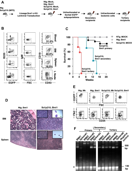
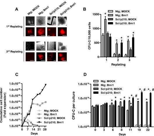
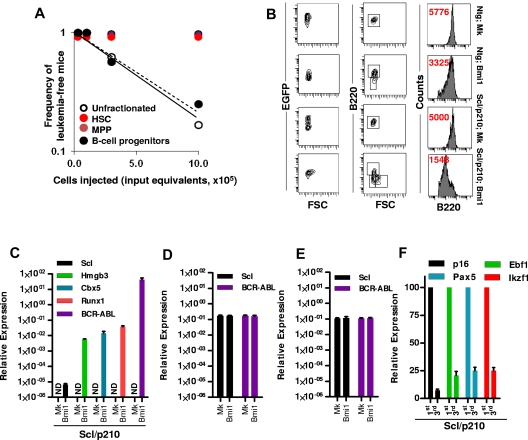
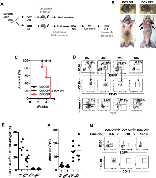
The characterization and targeting of Philadelphia chromosome positive (Ph(+)) acute lymphoblastic leukemia (ALL)-initiating cells remains unresolved. Expression of the polycomb protein Bmi1 is up-regulated in patients with advanced stages of chronic myelogenous leukemia (CML). We report that Bmi1 transforms and reprograms CML B-lymphoid progenitors into stem cell leukemia (Scl) promoter-driven, self-renewing, leukemia-initiating cells to result in B-lymphoid leukemia (B-ALL) in vivo. In vitro, highly proliferating and serially replatable myeloid and lymphoid colony-forming cultures could be established from BCR-ABL and Bmi1 coexpressing progenitors. However, unlike in vivo expanded CML B-lymphoid progenitors, hematopoietic stem cells, or multipotent progenitors, coexpressing BCR-ABL and Bmi1 did not initiate or propagate leukemia in a limiting dilution assay. Inducible genetic attenuation of BCR-ABL reversed Bmi1-driven B-ALL development, which was accompanied by induction of apoptosis of leukemic B-lymphoid progenitors and by long-term animal survival, suggesting that BCR-ABL is required to maintain B-ALL and that BCR-ABL and Bmi1 cooperate toward blast transformation in vivo. Our data indicate that BCR-ABL targeting itself is required to eradicate Ph(+)/Bmi1(+) B-ALL-initiating cells and confirm their addiction to BCR-ABL signaling.


Figures

Figure 1
Figure 1
Bmi1 collaborates with inducible BCR-ABL to induce lymphoid BC in vivo. (A) Experimental setup. To model CML-BC–initiating cells, LSK cells were isolated from Ntg or Scl/p210 mice presenting with myeloproliferative disorder (MPD), transduced with Bmi1 or empty lentiviral vectors expressing EGFP, and serially transplanted into lethally irradiated primary, secondary, and tertiary recipient mice. (B) Representative flow cytometry (FACS) contour diagram showing the frequency of EGFP+FSChiB220loCD19+CD43+IgM B-lymphoid blasts present in the peripheral blood, BM, spleen, and lymph nodes of Scl/p210;Bmi1 mice. (C) Cumulative survival using the Kaplan-Meier log-rank P test performed in Ntg;MOCK-, Ntg;Bmi1-, Scl/p210;MOCK-, and Scl/p210;Bmi1-transplanted primary, secondary, and tertiary recipient mice (P < .05, n = 6-12 mice per group; n = 2 independent experiments). (D) Histological evidence of leukemic cell infiltration into the BM (magnification 100×) and spleen (Magnification 100×) in Scl/p210;Bmi1 (DOX OFF) mice compared with Scl/p210;Bmi1 (DOX ON) mice. Inset pictures show tissue infiltration at higher magnification (400×) or cytospin preparation of immature lymphoblasts (magnification 1000×). (E) Representative FACS contour diagram showing the frequency of HSCs and MPPs gated on EGFP+ BM cells from Ntg;MOCK, Ntg;Bmi1, Scl/p210;MOCK, and Scl/p210;Bmi1 mice. (F) LAM-PCR amplifying lentiviral vector insertion sites in the BM and spleen of Scl/p210;Bmi1-transplanted primary, secondary, and tertiary recipient mice, demonstrating a predominantly monoclonal integration pattern. Negative control indicates mouse genomic DNA control. *Internal standard. Microphotographs were obtained with an Olympus CKX41, objectives ×10, ×40, and ×100. The images were acquired with a Moticam 2500 color camera (5.0 MPixal, USB2.0), Motich China Group Co Ltd, and processed using Motic Images Plus 2.0 software.
Figure 2
Figure 2
Bmi1 augments proliferation and maintains clonogenicity of Scl/p210 cells. (A) Representative fluorescence photographs showing CFU-Cs of Ntg;Mk-, Ntg;Bmi1-, Scl/p210;Mk-, and Scl/p210;Bmi1-expressing cells during serial replating on methylcellulose in vitro. Earlier, Bmi1 was subcloned into Sfβ91-IRES-RFP retroviral vector. HSC/Ps were transduced and RFP+ cells were sorted by flow cytometry (FACSAria II; BD Biosciences). (B) Frequency of CFU-Cs in the BM of Ntg;Mk-, Ntg;Bmi1-, Scl/p210;Mk-, and Scl/p210;Bmi1-expressing cells during serial replating on methylcellulose in vitro. (C) Cumulative cell number expansion of Ntg;Mk-, Ntg;Bmi1-, Scl/p210;Mk-, and Scl/p210;Bmi1-expressing cells grown in IMDM-based liquid culture supplemented with 10% FCS, 100 ng/mL of SCF, 100 ng/mL of thrombopoietin, and 100 ng/mL of G-CSF. Data represent 1 of 2 independent experiments with similar results. (D) CFU-C content (expansion) per culture of sorted RFP+ BM cells from Ntg;Mk-, Ntg;Bmi1-, Scl/p210;Mk-, and Scl/p210;Bmi1-expressing groups during liquid culture in respective time points in vitro. The CFU-C assay was performed in triplicate. Data represent 1 of 2 independent experiments with similar results. Microphotographs were obtained with a DC Imaging microscope, model BA310, with a ×20 objective (OM, ×200). The images were acquired with a Moticam 2500 color camera (5.0 MPixal, USB2.0), Motich China Group Co Ltd, and processed using Motich Image Plus 2.0 software.
Figure 3
Figure 3
Bmi-1 induces self-renewal and transforms B-lymphoid progenitors. (A) Frequency of leukemia-initiating cells in Scl/p210;Bmi1 mice in unfractionated and sorted cell fractions. Results are presented as cell input equivalents (P < .05, n = 8-10 mice per group). (B) Representative FACS contour diagram and histogram showing expression (in red, mean fluorescence intensity, MFI) of B220 on EGFP+ cells in Ntg;MOCK, Ntg;Bmi1, Scl/p210;MOCK, and Scl/p210;Bmi1 mice. (C) Relative mRNA expression (quantitative RT-PCR, 2ΔCt) analysis of Scl, Hmgb3, Cbx5, Runx1, and BCR-ABL (b3a2) in EGFP+ B-lymphoid progenitors isolated from Scl/p210;Mock (Mk) and Scl/p210;Bmi1 mice. ND indicates not detected. (D) Relative mRNA expression (quantitative RT-PCR, 2ΔCt) analysis of Scl, and BCR-ABL (b3a2) in EGFP+ HSCs isolated from Scl/p210;Mock (Mk) and Scl/p210;Bmi1 mice. (E) Relative mRNA expression (quantitative RT-PCR, 2ΔCt) analysis of Scl, and BCR-ABL (b3a2) in EGFP+ MPPs isolated from Scl/p210;Mock (Mk) and Scl/p210;Bmi1 mice. (F) Relative mRNA expression (quantitative RT-PCR, normalized) analysis of p16, Ebf1, Pax5, and Ikzf1 performed on EGFP+ B-lymphoid progenitors isolated from primary (first) and tertiary (third) Scl/p210;Bmi1 mice. Data were normalized to the leukemic cells obtained from primary (first) recipient mice.
Figure 4
Figure 4
BCR-ABL expression is indispensable for survival, initiation, and maintenance of Bmi1-expressing CML BC–initiating cells in vivo. (A) Experimental setup. To study leukemia initiation, mice transplanted with B-ALL–initiating cells were fed with (DOX ON) or without (DOX OFF) doxycycline immediately after transplantation. To study leukemia maintenance, recipient mice that had already developed B-ALL under DOX OFF conditions were switched back to a DOX ON diet to stop the further expression of BCR-ABL. (B) Representative image of Scl/p210;Bmi1 mice fed DOX-ON or DOX-OFF diet. (C) Cumulative survival of Scl/p210;Bmi1 mice during leukemia initiation and maintenance DOX-ON or DOX-OFF diets. (P < .05, n = 3-4 mice per group). (D-G) Mice fed with a DOX OFF followed by a DOX ON diet. (D) Representative FACS contour diagram showing EGFP+ B-lymphoid progenitors and annexin V+ B-lymphoid progenitors present in the peripheral blood of Scl/p210;Bmi1 mice. (E) Frequency of EGFP+ B-lymphoid progenitors present in the peripheral blood of Scl/p210;Bmi1 mice (n = 6 mice in each time point). (F) Frequency of annexin V+ B-lymphoid progenitors present in the peripheral blood of Scl/p210;Bmi1 mice (n = 6 mice in each time point). (G) Representative FACS contour diagram showing EGFP+ B-lymphoid progenitors present in the BM of Scl/p210;Bmi1 mice.

Similar articles

Cited by

References

    1. Notta F, Mullighan CG, Wang JC, et al. Evolution of human BCR-ABL1 lymphoblastic leukaemia-initiating cells. Nature. 2011;469(7330):362–367. - PubMed
    1. Smith KS, Chanda SK, Lingbeek M, et al. Bmi-1 regulation of INK4A-ARF is a downstream requirement for transformation of hematopoietic progenitors by E2a-Pbx1. Mol Cell. 2003;12(2):393–400. - PubMed
    1. Mullighan CG, Miller CB, Radtke I, et al. BCR-ABL1 lymphoblastic leukaemia is characterized by the deletion of Ikaros. Nature. 2008;453(7191):110–114. - PubMed
    1. Mullighan CG, Zhang J, Kasper LH, et al. CREBBP mutations in relapsed acute lymphoblastic leukaemia. Nature. 2011;471(7337):235–239. - PMC - PubMed
    1. Castor A, Nilsson L, Astrand-Grundstrom I, et al. Distinct patterns of hematopoietic stem cell involvement in acute lymphoblastic leukemia. Nat Med. 2005;11(6):630–637. - PubMed

Publication types

MeSH terms