. 2011 Nov 20;18(12):1388-93.
doi: 10.1038/nsmb.2168.

Mechanistic insights into the activation of oncogenic forms of EGF receptor

Affiliations

Mechanistic insights into the activation of oncogenic forms of EGF receptor

Zhihong Wang et al. Nat Struct Mol Biol. .

Abstract

Epidermal growth factor receptor (EGFR) is a receptor tyrosine kinase that is commonly activated by mutation in non-small cell lung cancer. The mechanism of this oncogenic activation is not completely understood, but in contrast to that of the wild-type EGFR, it is proposed to be independent of kinase domain dimerization. Mechanistic studies on EGFR have mainly relied on cell-based assays or isolated kinase domain measurements. Here we show, using purified, near full-length human EGFR proteins (tEGFRs), that two oncogenic mutants are fully active independently of EGF and highly resistant to the therapeutic and endogenous inhibitors cetuximab, lapatinib and MIG6. Based on the pattern of inhibition and the effects of additional asymmetric kinase dimer interface mutations, we propose that these oncogenic EGFR mutants drive and strongly depend on the formation of the asymmetric kinase dimer for activation, which has implications for drug design and cancer treatment strategies.


Figures
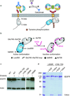
Figure 1
Figure 1. Activation and inhibition mechanism for WT EGFR and the expression and purification strategy for mutant tEGFRs
(a) Unliganded and Cetuximab–bound WT EGFR exist primarily in the tethered conformation. EGF binding to the ectodomain initiates formation of specific receptor-mediated dimers and activation of the intracellular kinase domain via formation of an asymmetric dimer. The active conformation of kinase domain is depicted as blue and the inactive conformation is depicted as gray. Cetuximab is shown in light blue and EGF is shown in purple. Not to scale. (b) MIG6 inhibits WT EGFR by binding to the C-lobe of the EGFR kinase domain and blocking the asymmetric dimer interface. Sites of key residues studied here are highlighted. (c) Western blot analysis of the expression levels of WT, L858R, and Δ746–750 tEGFRs in the presence and absence of the EGFR inhibitor erlotinib. HEK293 GnTi cells were transfected with the plasmid DNA encoding tEGFR, and cultured in the presence and absence of 50 nM erlotinib. (d) Coomassie blue-stained SDS-PAGE analysis of the purified L858R tEGFR and Δ746–750 tEGFR with either EGF or Cetuximab (Cetux) as ligand.
Figure 2
Figure 2. MIG6 seg 1+2 interacts with WT and mutant tEGFRs
(a) Autoradiograph showing incorporation of radioactive phosphate (32P) into tEGFR and MIG6 simultaneously by in vitro phosphorylation of MIG6 seg 1+2 (77 aa) using various tEGFR forms. MIG6 seg 1+2 was incubated with [32P] ATP and WT–EGF, L858R–EGF, L858R–Cetux, Δ(746–750)–EGF, and Δ(746–750)–Cetux tEGFRs. Left, negative control in which no MIG6 seg 1+2 was added to the reaction buffer. The relative intensity of each tEGFR band (divided by the intensity of the EGFR band on WT–EGF lane from each autoradiograph) is shown below. (b) MIG6 seg 1+2 is phosphorylated on tyrosine by tEGFRs in vitro. MIG6 seg 1+2 was incubated with ATP in the presence and absence of tEGFRs (WT tEGFR–EGF, L858R tEGFR–EGF, L858R tEGFR–Cetux, Δ(746–750) tEGFR–EGF, and Δ(746–750) tEGFR–Cetux). The tyrosine phosphorylation of MIG6 seg 1+2 was probed with anti-pY 4G10 antibody (upper panel). The coomassie staining of MIG6 seg 1+2 (lower panel) indicates that the total amount of MIG6 from each lane was identical. (c) The inhibition effects of MIG6 seg 1+2 on WT–EGF, WT kinase domain (residues 668–1210), L858R–EGF, L858R–Cetux, Δ(746–750)–EGF, and Δ(746–750)–Cetux tEGFRs. Percent of activity values (± s.d.) calculated from three independent experiments are shown.
Figure 3
Figure 3. Role of the asymmetric dimer interface for oncogenic EGFR activation
(a) Comparison of the specific activities of L858R–EGF, L858R I706Q–EGF, and L858R V948R–EGF tEGFRs. (b) Comparison of the specific activities of L858R–Cetux, L858R I706Q–Cetux, and L858R V948R–Cetux tEGFRs. (c) Comparison of the specific activities of Δ(746–750)–EGF, Δ(746–750) I706Q–EGF, and Δ(746–750) I706Q–Cetux tEGFRs. (d) Activation mechanism for EGFR oncogenic mutations. The unliganded mutant EGFR is present in the super-activated dimer conformation which is driven by kinase domain mutation (L858R shown or Δ746–750, not shown) and independent of ectodomain occupancy.

Comment in

  • Finding the missing links in EGFR.
    Bessman NJ, Lemmon MA. Bessman NJ, et al. Nat Struct Mol Biol. 2012 Jan 5;19(1):1-3. doi: 10.1038/nsmb.2221. Nat Struct Mol Biol. 2012. PMID: 22218287 No abstract available.

References

    1. Schlessinger J. Cell signaling by receptor tyrosine kinases. Cell. 2000;103:211–225. - PubMed
    1. Hynes NE, Lane HA. ERBB receptors and cancer: the complexity of targeted inhibitors. Nat Rev Cancer. 2005;5:341–354. - PubMed
    1. Wheeler DL, Dunn EF, Harari PM. Understanding resistance to EGFR inhibitors-impact on future treatment strategies. Nat Rev Clin Oncol. 2010;7:493–507. - PMC - PubMed
    1. Stamos J, Sliwkowski MX, Eigenbrot C. Structure of the epidermal growth factor receptor kinase domain alone and in complex with a 4-anilinoquinazoline inhibitor. J Biol Chem. 2002;277:46265–46272. - PubMed
    1. Lynch TJ, et al. Activating mutations in the epidermal growth factor receptor underlying responsiveness of non-small-cell lung cancer to gefitinib. N Engl J Med. 2004;350:2129–2139. - PubMed

Publication types