

doi: 10.1107/S1744309111029654.
Epub 2011 Sep 6.
Structure of New Delhi metallo-β-lactamase 1 (NDM-1)
Affiliations
- PMID: 22102018
- PMCID: PMC3212353
- DOI: 10.1107/S1744309111029654
Item in Clipboard
Structure of New Delhi metallo-β-lactamase 1 (NDM-1)
Acta Crystallogr Sect F Struct Biol Cryst Commun.
.
Abstract
Antibiotic resistance in bacterial pathogens poses a serious threat to human health and the metallo-β-lactamase (MBL) enzymes are responsible for much of this resistance. The recently identified New Delhi MBL 1 (NDM-1) is a novel member of this family that is capable of hydrolysing a wide variety of clinically important antibiotics. Here, the crystal structure of NDM-1 from Klebsiella pneumoniae is reported and its structure and active site are discussed in the context of other recently deposited coordinates of NDM-1.
© 2011 International Union of Crystallography. All rights reserved.
Figures
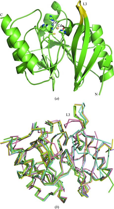
(a) The structure of NDM-1 shows that the protein contains the αβ/βα sandwich common to all MBLs despite the low sequence identity between NDM-1 and other metallo-β-lactamases. The metal-chelating residues within the active site are displayed as sticks. The zinc ion is coloured blue and the cadmium ion is coloured fawn. Flexible loop L3, which is proposed to play a role in substrate binding, is highlighted in yellow. (b) Superposition of our coordinates (green) with other models of NDM-1 (PDB entries 3q6x , cyan; 3rkj , violet; 3rkk , yellow; 3s0z , pink) shows the protein structure to be rather rigid with one key region of flexibility, loop L3. All figures were produced using PyMOL (Schrödinger LLC).
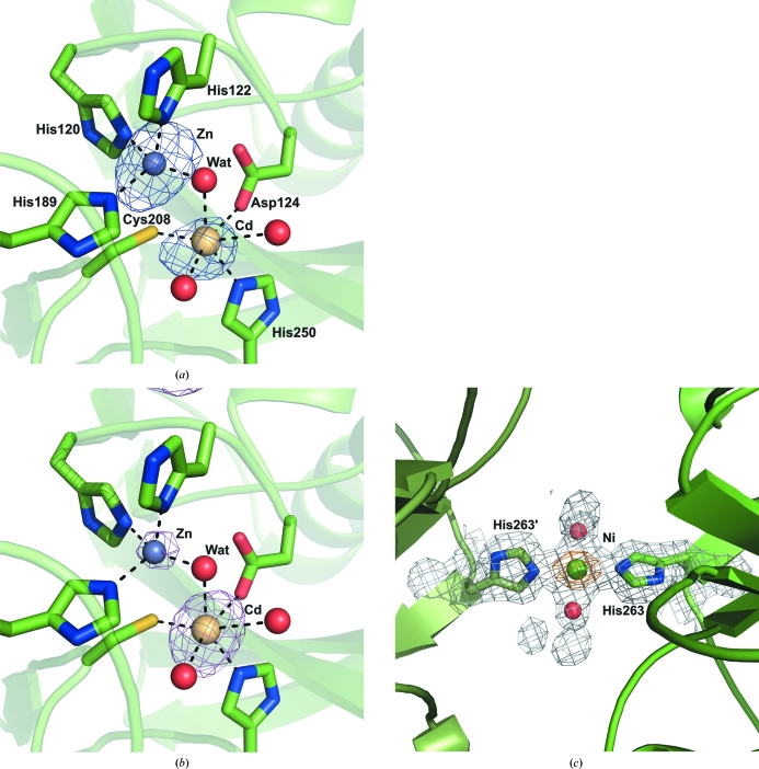
(a) The active site of NDM-1 contains a zinc ion (blue) in site 1 and a cadmium ion (fawn) in site 2, both of which are coordinated by the water molecule (labelled Wat) responsible for hydrolysis of the antibiotic. An anomalous difference Fourier map calculated using data collected at the Zn K edge shows a much larger peak for the metal ion in site 1 relative to that in site 2. The anomalous maps shown are all contoured at a level of 0.05 e (5σ). (b) An anomalous difference Fourier map calculated using data collected at the Co K edge shows a stronger peak for the metal ion at site 2. The relative peak heights are consistent with the assignment of the metal ions in the active site and at crystal contacts. (c) An example of a nickel ion (green) located at a crystal contact between His263 and the equivalent amino acid, labelled His263′, in an adjacent protein molecule. The anomalous difference map calculated using data collected at the Ni K edge is shown in orange.
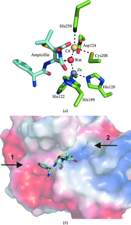
(a) An ampicillin molecule modelled into the active site of NDM-1 shows that the shared catalytic water molecule is ideally positioned to attack the β-lactam carbonyl group, as indicated by the arrow. (b) A representation of the van der Waals surface of NDM-1 shows that the substrate-binding groove on both sides of the metal ions is large enough to accommodate sizeable ligands, suggesting that β-lactam antibiotics carrying very large substituent groups could still be bound and hydrolysed by NDM-1. The point of variation between the penicillin-derived β-lactams is indicated by arrow 1 and the main point of variation in the carbepenem antibiotics is shown by arrow 2, both of which point into the large substrate-binding groove.
References
Publication types
MeSH terms
Substances
Associated data
- Actions
Grants and funding
LinkOut - more resources
Full Text Sources
Other Literature Sources
Miscellaneous
