. 2011 Nov 1;67(Pt 11):1316-22.
doi: 10.1107/S1744309111030417. Epub 2011 Oct 25.

Novel crystallization conditions for tandem variant R67 DHFR yield a wild-type crystal structure

Affiliations

Novel crystallization conditions for tandem variant R67 DHFR yield a wild-type crystal structure

Brahm J Yachnin et al. Acta Crystallogr Sect F Struct Biol Cryst Commun. .

Abstract

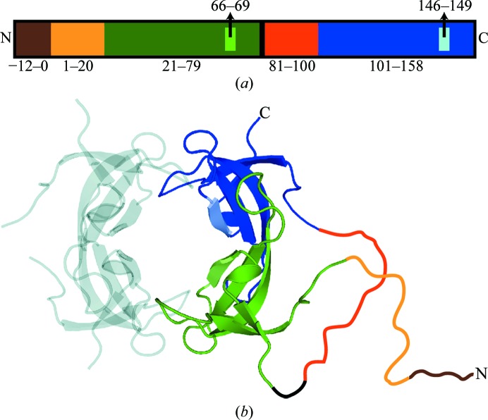
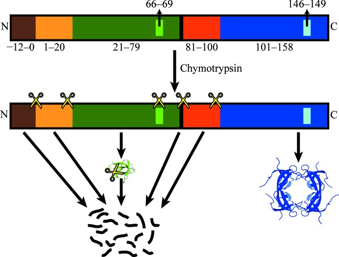
Trimethoprim is an antibiotic that targets bacterial dihydrofolate reductase (DHFR). A plasmid-encoded DHFR known as R67 DHFR provides resistance to trimethoprim in bacteria. To better understand the mechanism of this homotetrameric enzyme, a tandem dimer construct was created that linked two monomeric R67 DHFR subunits together and mutated the sequence of residues 66-69 of the first subunit from VQIY to INSF. Using a modified crystallization protocol for this enzyme that included in situ proteolysis using chymotrypsin, the tandem dimer was crystallized and the structure was solved at 1.4 Å resolution. Surprisingly, only wild-type protomers were incorporated into the crystal. Further experiments demonstrated that the variant protomer was selectively degraded by chymotrypsin, although no canonical chymotrypsin cleavage site had been introduced by these mutations.


Figures

Figure 1
Figure 1
Organization of the INSF tandem dimer gene. (a) The different segments of the primary structure of the INSF tandem dimer are shown to scale. The colours represent the various segments of the gene. Brown (residues −12 to 0) represents the His tag, light orange (residues 1–20) and orange (residues 81–100) represent the N-terminal unstructured regions in the variant and wild-type protomers, respectively, green (residues 21–78) and blue (residues 101–158) represent the structured regions in the variant and wild-type protomers, respectively, and black (residues 79–80) represents the interprotomer linker. Lighter green (residues 66–69) and blue (residues 146–149) represent the mutation sites in the variant (INSF) and wild-type (VQIY) protomers, respectively. (b) The same colour scheme is used to show the proposed structure of the tandem dimer in the context of the assembled tetramer. The positions of the unstructured regions and linker (drawn in) are speculated.
Figure 2
Figure 2
Change in magnitude of map σ and R factor with increasing wild-type character. The map σ value is evaluated at the position of one atom per residue that is unique to either the wild-type or the variant protomers. The map σs (left axis) that are positive are shown in green and denoted with a + sign, while the map σs that are negative are shown in red with a − sign. R factors (right axis) are shown in blue.
Figure 3
Figure 3
Electron-density map of the mutation sites. 2F oF c (blue) and F oF c (green and red) maps contoured at a map σ of 1.5 and 3.0, respectively, calculated using (a) the wild-type and (b) the variant models are shown. The wild-type model is shown in green and the variant model is shown in brown. The model used to calculate the map in each panel is shown using thicker lines.
Figure 4
Figure 4
Comparison of the untreated and chymotrypsin-treated INSF tandem dimers on SDS–PAGE. The positions of the uncleaved (tandem dimer) and digested monomeric proteins are shown and the expected molecular weight as determined by mass spectroscopy is given.
Figure 5
Figure 5
Proposed model for the degradation of the INSF tandem dimer by chymotrypsin. Proposed chymotrypsin cut sites are denoted using the scissors icon. The exact positions are speculative and there may be additional cut sites that are not shown. The colour scheme is the same as is used in Fig. 1 ▶.

References

    1. Bradrick, T. D., Beechem, J. M. & Howell, E. E. (1996). Biochemistry, 35, 11414–11424. - PubMed
    1. DeLano, W. L. (2002). PyMOL http://www.pymol.org.
    1. Divya, N., Grifith, E. & Narayana, N. (2007). Protein Sci. 16, 1063–1068. - PMC - PubMed
    1. Dong, A. et al. (2007). Nature Methods, 4, 1019–1021. - PMC - PubMed
    1. Emsley, P. & Cowtan, K. (2004). Acta Cryst. D60, 2126–2132. - PubMed

Publication types

Substances

Associated data