. 2011 Nov 1;67(Pt 11):1339-44.
doi: 10.1107/S1744309111032507. Epub 2011 Oct 25.

Structure of the catalytic domain of Plasmodium falciparum ARF GTPase-activating protein (ARFGAP)

Affiliations

Structure of the catalytic domain of Plasmodium falciparum ARF GTPase-activating protein (ARFGAP)

William J Cook et al. Acta Crystallogr Sect F Struct Biol Cryst Commun. .

Abstract

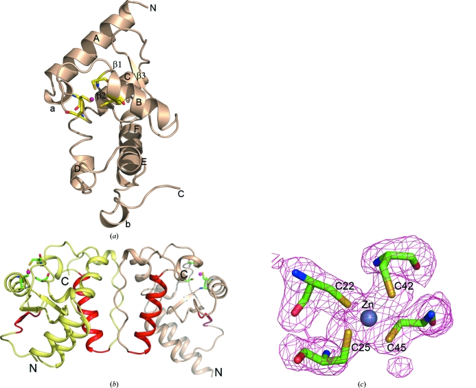
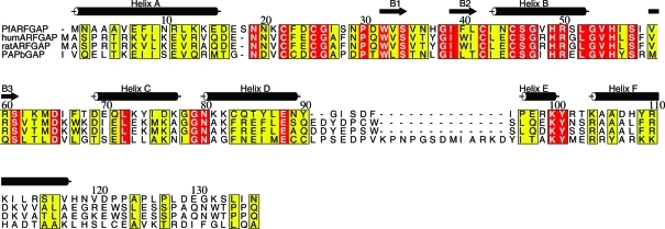
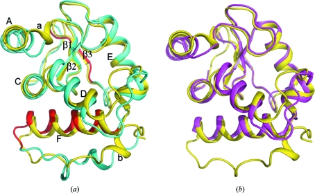
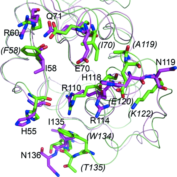
The crystal structure of the catalytic domain of the ADP ribosylation factor GTPase-activating protein (ARFGAP) from Plasmodium falciparum has been determined and refined to 2.4 Å resolution. Multiwavength anomalous diffraction (MAD) data were collected utilizing the Zn(2+) ion bound at the zinc-finger domain and were used to solve the structure. The overall structure of the domain is similar to those of mammalian ARFGAPs. However, several amino-acid residues in the area where GAP interacts with ARF1 differ in P. falciparum ARFGAP. Moreover, a number of residues that form the dimer interface in the crystal structure are unique in P. falciparum ARFGAP.


Figures

Figure 1
Figure 1
Structure of the catalytic domain of PfARFGAP. (a) Cartoon drawing of the structure of PfARFGAP. The four cysteine residues that form the zinc-binding site are shown as stick models. The Zn2+ ion is shown as a magenta sphere. The α-helices are labeled A–F and the β-strands are labeled β2 and β3; β1 remains at the back. The 310-helices are labeled a and b. (b) Cartoon drawing of the two molecules related by the noncrystallographic twofold axis in the asymmetric unit. The four cysteine residues that form the zinc-binding site in each molecule are shown as stick models. The Zn2+ ions are shown as magenta spheres. The residues involved in binding ARF, based on the ARF–ARFGAP structure (Goldberg, 1999 ▶), are shown in red. (c) Cysteine residues in the zinc-finger domain of molecule A with associated electron density. Figs. 1, 3, 4 and 5 were created with PyMOL (DeLano, 2002 ▶).
Figure 2
Figure 2
Alignment of the primary sequence of residues 1–136 of PfARFGAP with the corresponding residues of rat and human ARFGAPs and mouse PAPβGAP. Sequences are taken from GenBank accession Nos. AAN36512 (PfARFGAP), AAH00786 (human), AAH70895 (rat) and Q7SIG6 (mouse). Residue 8 in the PfARFGAP structure was found to be phenylalanine but was reported as leucine in the database. The α-helices and β-strands in PfARFGAP are indicated. Note that two additional short β-strands are assigned in the rat ARFGAP structure (Goldberg, 1999 ▶) but are not seen in PfARFGAP. This figure was prepared using ALSCRIPT (Barton, 1993 ▶).
Figure 3
Figure 3
Dimer interface. The stereo drawing illustrates interactions that stabilize the conformation of the interface between monomers in the asymmetric unit. Amino-acid residues are shown as stick models. Important hydrogen bonds are indicated by dashed lines. Several amino-acid residues at the interface are distinctive in the PfARFGAP sequence.
Figure 4
Figure 4
Alignment of structures. (a) Structures of the catalytic domain of PfARFGAP (yellow) and rat ARFGAP (cyan) are superimposed. The ARF-binding region, based on the ARF–rat ARFGAP structure (Goldberg, 1999 ▶), is colored red. α-Helices and β-strands in PfARFGAP are labeled A–F and 1–3, respectively; 310-helices are labeled a and b. (b) Structures of the catalytic domain of PfARFGAP (yellow) and human ARFGAP (magenta) are superimposed in the same orientation as in (a). Note that the human ARFGAP structure (PDB entry 3dwd) only extends to residue 120.
Figure 5
Figure 5
Potential ARF-binding regions. Comparison of the residues involved in binding ARF, based on the ARF–ARFGAP structure (Goldberg, 1999 ▶). Crystal structures of PfARFGAP (light magenta) and rat ARFGAP (green) are superimposed. The residues in rat ARFGAP (green) that differ from the corresponding residues in PfARFGAP (magenta) are indicated in italics and in parentheses.

References

    1. Barton, G. J. (1993). Protein Eng. 6, 37–40. - PubMed
    1. Baumgartner, F., Wiek, S., Paprotka, K., Zauner, S. & Lingelbach, K. (2001). Mol. Microbiol. 41, 1151–1158. - PubMed
    1. Bi, X., Corpina, R. A. & Goldberg, J. (2002). Nature (London), 419, 271–277. - PubMed
    1. Bonifacino, J. S. & Glick, B. S. (2004). Cell, 116, 153–166. - PubMed
    1. Brünger, A. T. (1992). Nature (London), 355, 472–475. - PubMed

Publication types

Substances

Associated data