

The cerebral representation of temporomandibular joint occlusion and its alternation by occlusal splints
- PMID: 22102437
- PMCID: PMC6870206
- DOI: 10.1002/hbm.21466
The cerebral representation of temporomandibular joint occlusion and its alternation by occlusal splints
Abstract
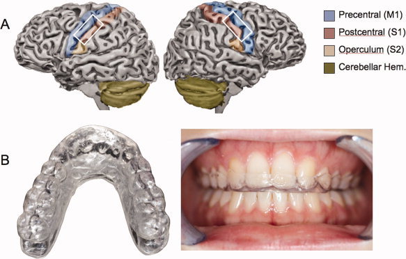
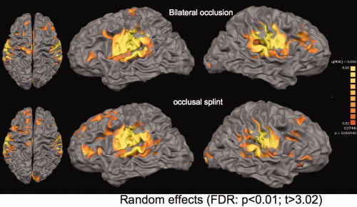
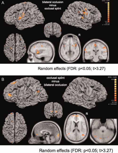
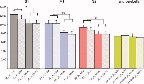
Occlusal splints are a common and effective therapy for temporomandibular joint disorder. Latest hypotheses on the impact of occlusal splints suggest an altered cerebral control on the occlusion movements after using a splint. However, the impact of using a splint during chewing on its cerebral representation is quite unknown. We used functional magnetic resonance imaging (fMRI) to investigate brain activities during occlusal function in centric occlusion on natural teeth or on occlusal splints in fifteen healthy subjects. Comparisons between conditions revealed an increased activation for the bilateral occlusion without a splint in bilateral primary and secondary sensorimotor areas, the putamen, inferior parietal and prefrontal cortex (left dorsal and bilateral orbital) and anterior insular. In contrast, using a splint increased activation in the bilateral prefrontal lobe (bilateral BA 10), bilateral temporo-parietal (BA 39), occipital and cerebellar hemispheres. An additionally applied individually based evaluation of representation sites in regions of interest demonstrated that the somatotopic representation for both conditions in the pre- and postcentral gyri did not significantly differ. Furthermore, this analysis confirmed the decreasing effect of the splint on bilateral primary and secondary motor and somatosensory cortical activation. In contrast to the decreasing effect on sensorimotor areas, an increased level of activity in the fronto-parieto-occipital and cerebellar network might be associated with the therapeutic effect of occlusal splints.
Copyright © 2011 Wiley Periodicals, Inc.
Figures
References
-
- Binkofski F, Buccino G, Posse S, Seitz RJ, Rizzolatti G, Freund H ( 1999): A fronto‐parietal circuit for object manipulation in man: Evidence from an fMRI‐study. Eur J Neurosci 11: 3276–3286. - PubMed
-
- Botelho AL, Silva BC, Gentil FH, Sforza C, da Silva MA ( 2010): Immediate effect of the resilient splint evaluated using surface electromyography in patients with TMD. Cranio 28: 266–273. - PubMed
-
- Carlsson GE ( 2009): Critical review of some dogmas in prosthodontics. J Prosthodont Res 53: 3–10. - PubMed
-
- Craig AD ( 2002): How do you feel? Interoception: The sense of the physiological condition of the body. Nat Rev Neurosci 3: 655–666. - PubMed
MeSH terms
LinkOut - more resources
Full Text Sources
Medical
