. 2012 Mar;40(5):2224-33.
doi: 10.1093/nar/gkr1001. Epub 2011 Nov 18.

Cleavage mediated by the P15 domain of bacterial RNase P RNA

Affiliations

Cleavage mediated by the P15 domain of bacterial RNase P RNA

Ema Kikovska et al. Nucleic Acids Res. 2012 Mar.

Abstract

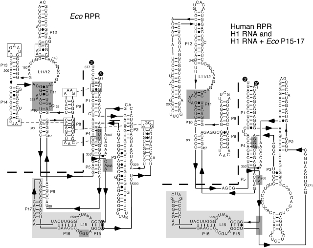
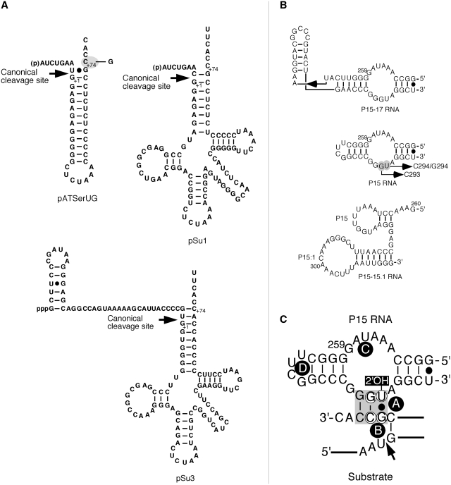
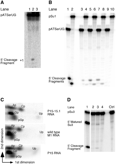
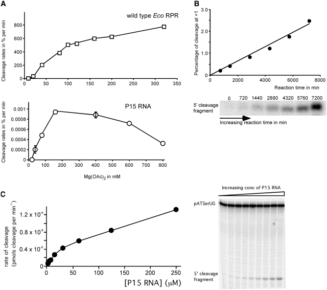
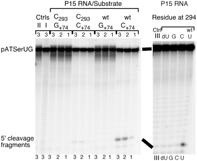
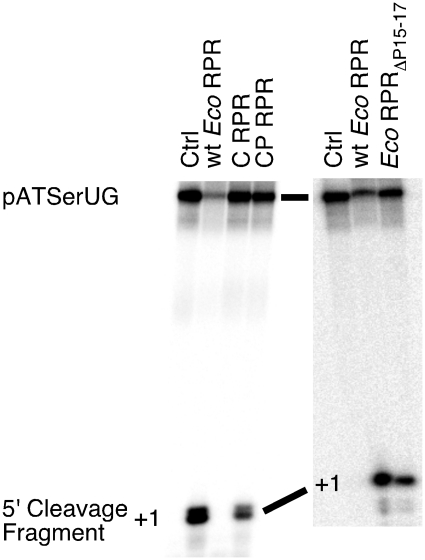
Independently folded domains in RNAs frequently adopt identical tertiary structures regardless of whether they are in isolation or are part of larger RNA molecules. This is exemplified by the P15 domain in the RNA subunit (RPR) of the universally conserved endoribonuclease P, which is involved in the processing of tRNA precursors. One of its domains, encompassing the P15 loop, binds to the 3'-end of tRNA precursors resulting in the formation of the RCCA-RNase P RNA interaction (interacting residues underlined) in the bacterial RPR-substrate complex. The function of this interaction was hypothesized to anchor the substrate, expose the cleavage site and result in re-coordination of Mg(2+) at the cleavage site. Here we show that small model-RNA molecules (~30 nt) carrying the P15-loop mediated cleavage at the canonical RNase P cleavage site with significantly reduced rates compared to cleavage with full-size RPR. These data provide further experimental evidence for our model that the P15 domain contributes to both substrate binding and catalysis. Our data raises intriguing evolutionary possibilities for 'RNA-mediated' cleavage of RNA.


Figures

Figure 1.
Figure 1.
Predicted secondary structures of wild-type Eco RPR and human RPR, H1 RNA (58). The specificity (S) and catalytic (C) domains are separated with the dashed line and the Eco RPR P15–P17 domain is highlighted in light grey. The Eco RPR P15–P17 domain was introduced into H1 RNA generating H1 RNA + Eco P15–P17 as indicated at the position marked with a grey box. Eco RPRΔP15–P17 carries a deletion of P15–P17 except the GU pair marked with black circles (22). The highlighted regions in dark grey mark regions in RPR (including P15–P17) known to be important for binding of the substrate and that show structural differences, for details see main text. P refers to helices in Eco RPR and H1 RNA.
Figure 2.
Figure 2.
(A) Secondary structures of pATSerUG, pSu1 and pSu3. The canonical RNase P cleavage sites are marked with arrows. The 3′-CCA residues in pATSerUG correspond to positions +74, +75 and +76 in full-length tRNA precursors and consequently the numbering of these positions follows the numbering in the tRNA precursors. The residue C+74 in pATSerUG was mutated to G as indicated. pSu1 and pSu3 correspond to the precursors to tRNASerSu1 and tRNATyrSu3, respectively. (B) The secondary structures of P15–P17 RNA, P15 RNA, and P15–P15.1 RNA. These RNAs were generated as outlined in ‘Materials and Methods’ section. P15–P17 RNA and P15 RNA were based on Eco RPR (Figure 1) while the P15–P15.1 RNA was based on M. hyopneumoniae RPR (31). The highlight residue marked in grey corresponds to G293 and was changed to C as indicated. (C) Model illustrating the interaction between the P15 RNA and the RCC-motif of the substrate (grey area), the RCCA–RPR interaction (interacting residues underlined). The residues in the P15 RNA that correspond to residues C293 and U294 in Eco RPR are encircled. These residues were replaced, C293 with G and U294 with G or C. The encircled residues in the substrate correspond to G+73 and C+74 in the substrate, and C+74 was substituted with G. The 2′-OH of U294 is highlighted and was replaced with 2′-H. The arrow marks the cleavage site and A–D (encircled in black) corresponds to Mg2+ ions that have been identified in the P15 RNA and in the substrate (19).
Figure 3.
Figure 3.
(A) Cleavage of pATSerUG with H1 RNA (lane 2) and H1P15–P17 RNA (lane 3). The concentrations of substrates and catalytic RNA were: ≤20 nM for the substrates, 4.8 µM for H1 RNA and 4.3 µM for H1P15–P17 RNA (Eco RPR P15–P17 inserted into H1 RNA; see Figure 1). Lane 1 pATSerUG incubated under the same conditions without RPR. Reaction times were 24 h in all cases. (B) Cleavage of pATSerUG and pSu1 with different RNAs. Lane 1, pATSerUG incubated with wild-type Eco RPR; lane 2, pSu1 incubated with wild-type Eco RPR; lane 3, pATSerUG incubated with P15 RNA; lane 4, pSu1 incubated with P15 RNA; lane 5, pATSerUG incubated with P15–P17 RNA; lane 6, pSu1 incubated with P15–P17 RNA; lane 7, pATSerUG incubated with P15–P15.1 RNA; lane 8, pSu1 incubated with P15–P15.1 RNA and lanes 9 and 10, incubation of pATSerUG and pSu1 alone, respectively. The concentrations of substrates and catalytic RNA were: ≤20 nM for the substrates, 0.16 µM for wild-type Eco RPR, 24 µM for P15–P17 RNA, 39 µM for P15 RNA and 23 µM for P15–P15.1 RNA. Reaction times were 20.5 h in all cases except for wild-type Eco RPR (10 sec). All reactions were performed at 160 mM Mg2+ in reaction buffer C at 37°C. (C) Two-dimensional TLC demonstrating the presence of pGp at the 5′-end of the 5′-matured cleavage product after cleavage of [α-32P]GTP internally labeled pATSerUG as indicated (14). (D) Cleavage of [α-32P]UTP internally labeled pSu3 (final specific activity ≥5 Ci/mmol) in reaction buffer C (see ‘Materials and Methods’ section) with different catalytic RNAs: lane 1 wild-type Eco RPR, lane 2 P15 RNA, lane 3 P15–P17 RNA and lane 4 P15–P15.1 RNA. The reaction time was 22 h in all cases except for wild-type Eco RPR (10 sec). The concentrations of: wild-type Eco RPR 0.16 µM, P15 RNA 39 µM, P15–P17 RNA 24 µM and P15–P15.1 23 µM. Negative control (Ctrl) incubation of pSu3 alone in reaction buffer C for 22 h. For further details see ‘Materials and Methods’ section.
Figure 4.
Figure 4.
(A) Cleavage of pATSerUG with wild-type Eco RPR and P15 RNA expressed as a percentage of cleavage per min as a function of Mg2+. Concentration of: substrate ≤20 nM, wild-type Eco RPR 3.2 µM and P15 RNA 39.1 µM. Data are the average of two independent experiments. Bars indicate the experimental range. (B) Cleavage of pATSerUG in percentage as a function of time and accumulation of the 5′-cleavage fragment over time as indicated. Same concentrations of P15 RNA and substrate as in Figure 3 were used. (C) A typical experiment illustrating cleavage of pATSerUG with P15 RNA as a function of increasing concentration of P15 RNA. The concentration of substrate was ≤20 nM and the reaction time after mixing of substrate and P15 RNA was 25 h.
Figure 5.
Figure 5.
Cleavage of different pATSerUG derivatives with chemically synthesized variants of the P15 RNA at 160 mM Mg2+ as indicated. The experiments were performed under single-turnover conditions at pH 6.0 and at 37°C as described in ‘Materials and Methods’ section. The concentrations of substrates and P15 RNA variants were ≤20 nM and 39.1 µM, respectively. In cleavage of the +74 variants the reaction times were 4 h (lanes labeled 1), 24.5 h (lanes labeled 2) and 26 h (lanes labeled 3). C+74 (wild-type pATSerUG) and G+74 refer to the identity of the residue at position 74 in the substrate while C293 refers to the identity of the residue at position 293 (wild-type: G293) in the P15 RNA (for comparison we use Eco RPR numbering; Figure 1A). In the right panel pATSerUG was cleaved with chemically synthesized P15 RNA variants carrying substitutions at position 294 (Figure 1C). A reaction time of 18 h was used while assaying the different P15 variants. Controls (Ctrl) incubation of substrate in reaction buffer C without the P15 RNA; Ctrl I pATSerUG (26 h); Ctrl II pATSerUG(G+74) (26 h); Ctrl III pATSerUG (18 h).
Figure 6.
Figure 6.
Cleavage activities of pATSerUG for the Eco RPR C domain with and without the P15–P17 domain (left panel), and for wt Eco RPR and Eco RPRΔP15–P17 (right panel). Amount of RNA added: C construct 7 µM and CP construct 5.5 µM. For wt Eco RPR and Eco RPRΔP15–P17, we used 0.8 µM and 2.8 µM, respectively. Irrespective of RPR construct the concentration of γ-32P 5′-end labeled pATSerUG was ≤20 nM. Time of incubations were: 4 h for the Eco RPR C-domain variants, 0.5 min for wt Eco RPR and 4 h for Eco RPRΔP15–P17. Ctrl (control): incubation of pATSerUG alone for 4 h in the reaction buffer C.

Similar articles

Cited by

References

    1. Masquida B, Westhof E. A modular and hierarchial approach for all-atom RNA modeling. In: Gesteland RF, Cech TR, Atkins JF, editors. The RNA World. 3rd edn. Cold Spring Harbor, NY: Cold Spring Harbor Press; 2006. pp. 659–681.
    1. Hendrix DK, Brenner SE, Holbrook SR. RNA structural motifs: building blocks of a molecular biomolecule. Q. Rev. Biophys. 2005;38:221–243. - PubMed
    1. Leontis NB, Lescoute A, Westhof E. The building blocks and motifs of RNA architecture. Curr. Opin. Struct. Biol. 2006;16:279–287. - PMC - PubMed
    1. Michel F, Westhof E. Modelling of the three-dimensional architecture of group I catalytic introns based on comparative sequence analysis. J. Mol. Biol. 1990;216:585–610. - PubMed
    1. Murphy FL, Cech TR. An independently folding domain of RNA tertiary structure withion the Tetrahymna ribozyme. Biochemistry. 1993;32:5291–5300. - PubMed

Publication types