. 2012 Feb;135(Pt 2):404-17.
doi: 10.1093/brain/awr288. Epub 2011 Nov 18.

Priming of adult pain responses by neonatal pain experience: maintenance by central neuroimmune activity

Affiliations

Priming of adult pain responses by neonatal pain experience: maintenance by central neuroimmune activity

Simon Beggs et al. Brain. 2012 Feb.

Abstract

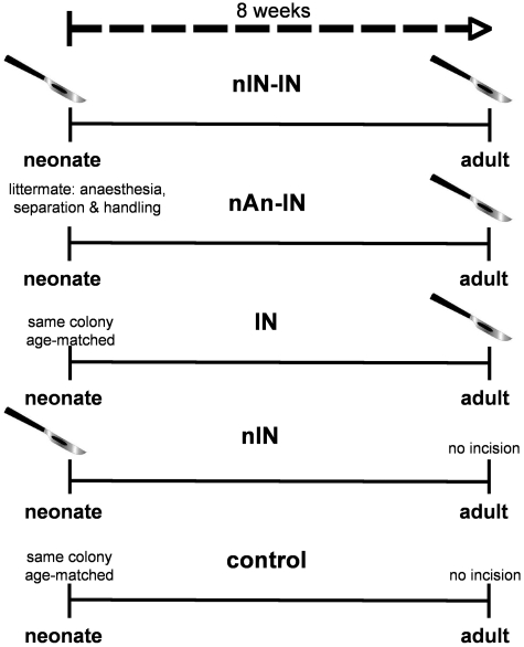
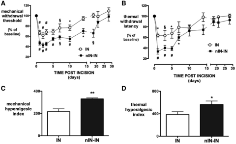
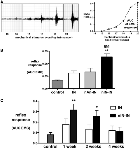
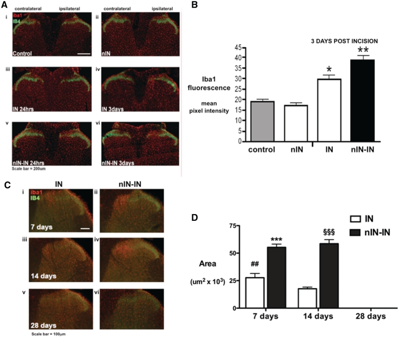
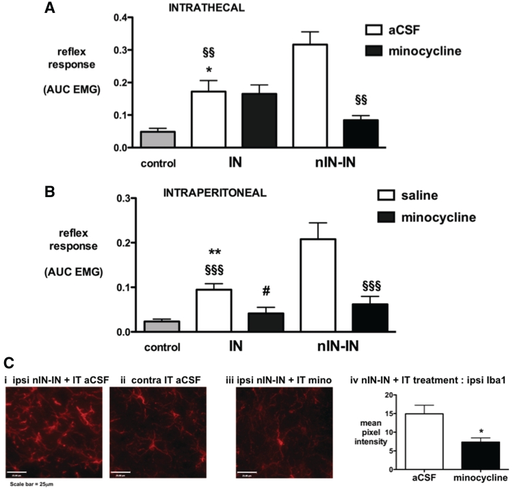
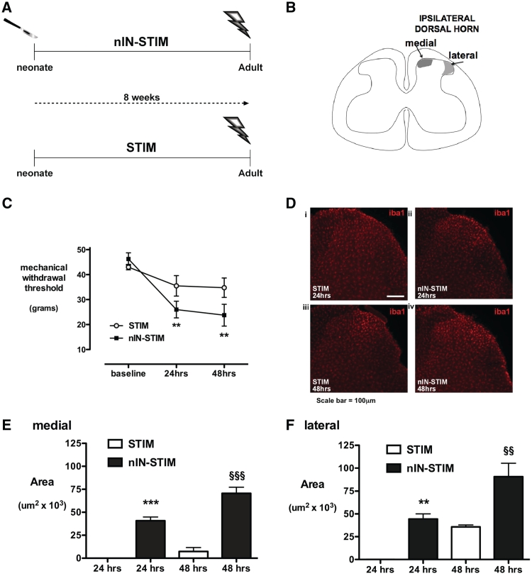
Adult brain connectivity is shaped by the balance of sensory inputs in early life. In the case of pain pathways, it is less clear whether nociceptive inputs in infancy can have a lasting influence upon central pain processing and adult pain sensitivity. Here, we show that adult pain responses in the rat are 'primed' by tissue injury in the neonatal period. Rats that experience hind-paw incision injury at 3 days of age, display an increased magnitude and duration of hyperalgesia following incision in adulthood when compared with those with no early life pain experience. This priming of spinal reflex sensitivity was measured by both reductions in behavioural withdrawal thresholds and increased flexor muscle electromyographic responses to graded suprathreshold hind-paw stimuli in the 4 weeks following adult incision. Prior neonatal injury also 'primed' the spinal microglial response to adult injury, resulting in an increased intensity, spatial distribution and duration of ionized calcium-binding adaptor molecule-1-positive microglial reactivity in the dorsal horn. Intrathecal minocycline at the time of adult injury selectively prevented both the hyperalgesia and early microglial reactivity associated with prior neonatal injury. The enhanced neuroimmune response seen in neonatally primed animals could also be demonstrated in the absence of peripheral tissue injury by direct electrical stimulation of tibial nerve fibres, confirming that centrally mediated mechanisms contribute to these long-term effects. These data suggest that early life injury may predispose individuals to enhanced sensitivity to painful events.


Figures

Figure 1
Figure 1
Schematic of experimental groups. Groups include: (i) nIN-IN: neonatal incision on post-natal Day 3 and repeat incision 8 weeks later in adulthood; (ii) nAn-IN: littermate control with equivalent anaesthesia, handling and maternal separation on post-natal Day 3 and incision in adulthood (post-natal Day 60); (iii) IN: age- matched animals from the same colony having incision in adulthood; (iv) nIN: animals having neonatal incision and follow-up in adulthood; and (iv) control: age-matched non-incised controls from the same colony (control).
Figure 2
Figure 2
Behavioural allodynia following injury in primed (nIN-IN) and non-primed (IN) adult animals. Ipsilateral hind-limb mechanical withdrawal thresholds (A) and thermal withdrawal latencies (B) are expressed as percentage change from baseline for 4 weeks following injury. Bars = mean ± SEM; n = 8 per group, 0–14 days; n = 4 per group, 18–28 days; and *P < 0.05, §P < 0.01 and #P ≤ 0.001, one-way repeated measures ANOVA with Dunnett's comparison with baseline; n = 4 per group, 0–28 days. The mechanical (C) and thermal (D) hyperalgesic index (area over the mechanical threshold or thermal latency curve for each animal for 14 days; area over the curve, 0–14 days) was also increased by neonatal priming. Bars = mean ± SEM; n = 8 per group; and *P < 0.05 and **P < 0.01, unpaired two-tailed Student's t-test.
Figure 3
Figure 3
Electromyographic quantification of injury-induced changes in reflex sensitivity. (A, left) Example trace of biceps femoris EMG responses to hind-paw mechanical stimuli. Von Frey hairs (number 15–20) deliver forces of 22, 37, 60, 90, 120 and 180 g, respectively. (A, right) The root mean square (RMS) of the EMG response is plotted against the mechanical stimulus and the area under the stimulus-response curve calculated to quantify the reflex response (AUC EMG). (B) The flexor reflex EMG response is shown in age-matched same colony controls (n = 12) and 24 h following incision in IN (age-matched same colony; n = 6), nAN-IN (littermates; n = 6) and in nIN-IN (n = 12) animals. Bars = mean ± SEM; **P < 0.01, nIN-IN versus nAn-IN and nIN-IN versus IN; §§§P < 0.001, nIN-IN versus control and one-way ANOVA with Bonferroni post hoc comparisons. (C) Flexor reflex EMG response 1, 2 and 4 weeks following injury in primed (nIN-IN) and non-primed (IN) age-matched adults. Bars = mean ± SEM; n = 5–6 per group; *P < 0.05 and **P < 0.01, one-way ANOVA with Dunnett's comparison with control.
Figure 4
Figure 4
Iba1 immunoreactivity in the dorsal horn of the spinal cord in primed and non-primed adults. (A) Lumbar spinal cord sections stained with Iba1 (red) and IB4 (green). Examples are shown from adult tissue: (i) control; (ii) neonatal incision only at post-natal Day 3 (nIN); (iii) 24 h following adult IN; (iv) 3 days following IN; (v) 24 h following nIN-IN; and (vi) 3 days following nIN-IN. Iba1 immunoreactivity is increased in the medial aspect of the superficial dorsal horn in nIN-IN at 24 h and 3 days, but only at 3 days in IN. (B) The intensity of Iba1 immunofluorescence in the same area of ipsilateral medial dorsal horn was quantified by integrated pixel density in non-incised controls (control), adults with neonatal incision only (nIN), and 3 days following incision in adults with (nIN-IN) and without (IN) neonatal incision. Bars = mean ± SEM; n = 4 animals per group; *P < 0.05 and **P < 0.01, one-way ANOVA with post hoc comparisons. (C) Spinal cord sections 7, 14 and 28 days following IN (i, iii and v) and nIN-IN (ii, iv and vi). (D) The area of the ipsilateral medial dorsal horn containing more than twice the density of Iba1 immunoreactivity than the equivalent contralateral region is increased 7 days following IN and at 7 and 14 days following nIN-IN. By 28 days there is no difference between ipsilateral and contralateral staining in either group (area of increased staining = 0). Bars = mean ± SEM; n = 4 animals per group, ##P < 0.01 IN versus baseline and ***P < 0.001, nIN-IN versus baseline and nIN-IN versus IN at 7 days; §§§P < 0.001, nIN-IN versus baseline and nIN-IN versus IN at 14 days; one-way ANOVA with Bonferroni post hoc comparisons.
Figure 5
Figure 5
Effects of minocycline on acute incision-related hyperalgesia. (A) The flexor reflex EMG response is shown in age-matched non-incised controls, and 24 h following incision in IN and nIN-IN groups that received intrathecal minocycline or artificial CSF (vehicle control). Bars = mean ± SEM; n = 6–8 per group; *P < 0.05, IN artificial CSF (aCSF) versus control, §§P < 0.01, nIN-IN artificial CSF versus IN artificial CSF and nIN-IN artificial CSF versus nIN-IN minocyline. (B) The flexor reflex EMG response is shown in age-matched non-incised controls, and 24 h following incision in IN and nIN-IN groups that received intraperitoneal minocycline or saline. Bars = mean ± SEM; n = 6–8 per group; **P < 0.01, IN saline versus control; and §§§P < 0.001, nIN-IN saline versus IN saline and nIN-IN minocycline. #P < 0.05, IN saline versus IN minocycline. (C) Representative high-power images from the medial dorsal horn of nIN-IN animals demonstrate increased Iba1 immunoreactivity in the (i) ipsilateral, but not (ii) contralateral side of the dorsal horn following intrathecal artificial CSF. Ipsilateral increases in Iba1 immunoreactivity are prevented by (iii) intrathecal minocycline and (iv) quantification of Iba1 immunofluorescence in the medial dorsal horn confirms a significant decrease in nIN-IN animals treated with intrathecal minocycline Bars = mean ± SEM; n = 4 per group; *P < 0.05, unpaired two-tailed Student's t-test.
Figure 6
Figure 6
Response to tibial nerve electrical stimulation at Aβ-, Aδ- and C-fibre intensity. (A) Treatment groups comprise: (i) nIN-STIM: neonatal incision (nIN) and tibial nerve stimulation (STIM) in adulthood; and (ii) STIM: tibial nerve stimulation in age-matched adults from the same colony. (B) Schematic of regions of increased microglial proliferation in the medial and lateral dorsal horn due to tibial stimulation and mid-thigh incision, respectively. (C) Mechanical withdrawal threshold at baseline, 24 and 48 h following tibial nerve electrical stimulation in nIN-STIM and STIM groups. Bars = mean ± SEM; n = 4 per group; **P < 0.01, one-way ANOVA with Dunnet's comparison with baseline. (D) Ipsilateral lumbar dorsal horn sections stained for Iba1 from STIM and nIN-STIM animals 24 (i and ii) and 48 h (iii and iv) following tibial nerve stimulation. The area of microglial proliferation in the medial (E) and lateral (F) dorsal horn is shown 24 and 48 h after tibial nerve electrical stimulation. At 24 h, Iba1 immunoreactivity is increased in the nIN-STIM but not the STIM group (area = 0), and to a greater degree in the nIN-STIM group at 48 h. Enhanced effects are seen in both the medial and lateral dorsal horn in neonatally primed animals (nIN-STIM). Bars = mean ± SEM; n = 4 animals each group; **P < 0.01 and ***P < 0.001, nIN-STIM versus STIM at 24 h; §§P < 0.01 and §§§P < 0.001, nIN-STIM versus STIM at 48 h; one-way ANOVA with Bonferroni post hoc comparisons.

References

    1. Alexander GM, van Rijn MA, van Hilten JJ, Perreault MJ, Schwartzman RJ. Changes in cerebrospinal fluid levels of pro-inflammatory cytokines in CRPS. Pain. 2005;116:213–9. - PubMed
    1. Banik RK, Woo YC, Park SS, Brennan TJ. Strain and sex influence on pain sensitivity after plantar incision in the mouse. Anesthesiology. 2006;105:1246–53. - PubMed
    1. Bastos LF, Merlo LA, Rocha LT, Coelho MM. Characterization of the antinociceptive and anti-inflammatory activities of doxycycline and minocycline in different experimental models. Eur J Pharmacol. 2007;576:171–9. - PubMed
    1. Beggs S, Salter MW. Stereological and somatotopic analysis of the spinal microglial response to peripheral nerve injury. Brain Behav Immun. 2007;21:624–33. - PMC - PubMed
    1. Beggs S, Salter MW. Microglia-neuronal signalling in neuropathic pain hypersensitivity 2.0. Curr Opin Neurobiol. 2010;20:474–80. - PMC - PubMed

Publication types