

Interaction of streptavidin-based peptide-MHC oligomers (tetramers) with cell-surface TCRs
- PMID: 22102724
- PMCID: PMC3237744
- DOI: 10.4049/jimmunol.1101734
Interaction of streptavidin-based peptide-MHC oligomers (tetramers) with cell-surface TCRs
Abstract
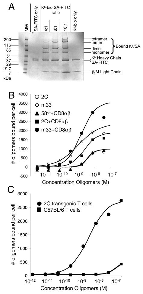
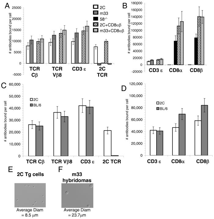
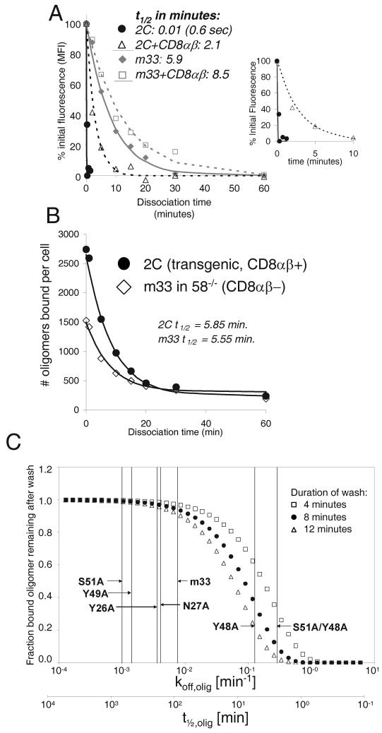
The binding of oligomeric peptide-MHC (pMHC) complexes to cell surface TCR can be considered to approximate TCR-pMHC interactions at cell-cell interfaces. In this study, we analyzed the equilibrium binding of streptavidin-based pMHC oligomers (tetramers) and their dissociation kinetics from CD8(pos) T cells from 2C-TCR transgenic mice and from T cell hybridomas that expressed the 2C TCR or a high-affinity mutant (m33) of this TCR. Our results show that the tetramers did not come close to saturating cell-surface TCR (binding only 10-30% of cell-surface receptors), as is generally assumed in deriving affinity values (K(D)), in part because of dissociative losses from tetramer-stained cells. Guided by a kinetic model, the oligomer dissociation rate and equilibrium constants were seen to depend not only on monovalent association and dissociation rates (k(off) and k(on)), but also on a multivalent association rate (μ) and TCR cell-surface density. Our results suggest that dissociation rates could account for the recently described surprisingly high frequency of tetramer-negative, functionally competent T cells in some T cell responses.
Figures
Similar articles
-
Functional evidence for TCR-intrinsic specificity for MHCII.Proc Natl Acad Sci U S A. 2016 Mar 15;113(11):3000-5. doi: 10.1073/pnas.1518499113. Epub 2016 Feb 1. Proc Natl Acad Sci U S A. 2016. PMID: 26831112 Free PMC article.
-
Interplay between T cell receptor binding kinetics and the level of cognate peptide presented by major histocompatibility complexes governs CD8+ T cell responsiveness.J Biol Chem. 2012 Jun 29;287(27):23068-78. doi: 10.1074/jbc.M112.357673. Epub 2012 May 1. J Biol Chem. 2012. PMID: 22549784 Free PMC article.
-
CD8(-) T cell transfectants that express a high affinity T cell receptor exhibit enhanced peptide-dependent activation.J Exp Med. 2001 Oct 15;194(8):1043-52. doi: 10.1084/jem.194.8.1043. J Exp Med. 2001. PMID: 11602635 Free PMC article.
-
The versatility of the αβ T-cell antigen receptor.Protein Sci. 2014 Mar;23(3):260-72. doi: 10.1002/pro.2412. Epub 2014 Jan 28. Protein Sci. 2014. PMID: 24375592 Free PMC article. Review.
-
A structural voyage toward an understanding of the MHC-I-restricted immune response: lessons learned and much to be learned.Immunol Rev. 2012 Nov;250(1):61-81. doi: 10.1111/j.1600-065X.2012.01159.x. Immunol Rev. 2012. PMID: 23046123 Review.
Cited by
-
Efficient magnetic enrichment of antigen-specific T cells by engineering particle properties.Biomaterials. 2018 Dec;187:105-116. doi: 10.1016/j.biomaterials.2018.09.029. Epub 2018 Sep 28. Biomaterials. 2018. PMID: 30312851 Free PMC article.
-
Monovalent engagement of the BCR activates ovalbumin-specific transnuclear B cells.J Exp Med. 2014 Feb 10;211(2):365-79. doi: 10.1084/jem.20131603. Epub 2014 Feb 3. J Exp Med. 2014. PMID: 24493799 Free PMC article.
-
Receptor Pre-Clustering and T cell Responses: Insights into Molecular Mechanisms.Front Immunol. 2014 Apr 30;5:132. doi: 10.3389/fimmu.2014.00132. eCollection 2014. Front Immunol. 2014. PMID: 24817867 Free PMC article. Review.
-
Deep Mutational Scans as a Guide to Engineering High Affinity T Cell Receptor Interactions with Peptide-bound Major Histocompatibility Complex.J Biol Chem. 2016 Nov 18;291(47):24566-24578. doi: 10.1074/jbc.M116.748681. Epub 2016 Sep 28. J Biol Chem. 2016. PMID: 27681597 Free PMC article.
-
Binding of TCR multimers and a TCR-like antibody with distinct fine-specificities is dependent on the surface density of HLA complexes.PLoS One. 2012;7(12):e51397. doi: 10.1371/journal.pone.0051397. Epub 2012 Dec 10. PLoS One. 2012. PMID: 23251518 Free PMC article.
References
-
- Sykulev Y, Brunmark A, Tsomides TJ, Kageyama S, Jackson M, Peterson PA, Eisen HN. High-affinity reactions between antigen-specific T-cell receptors and peptides associated with allogeneic and syngeneic major histocompatibility complex class I proteins. Proc Natl Acad Sci U S A. 1994;91:11487–11491. - PMC - PubMed
-
- Serbina N, Pamer EG. Quantitative studies of CD8+ T-cell responses during microbial infection. Curr Opin Immunol. 2003;15:436–442. - PubMed
Publication types
MeSH terms
Substances
Grants and funding
LinkOut - more resources
Full Text Sources
Other Literature Sources
Research Materials
