. 2011 Nov;7(11):e1002372.
doi: 10.1371/journal.ppat.1002372. Epub 2011 Nov 10.

Galactosaminogalactan, a new immunosuppressive polysaccharide of Aspergillus fumigatus

Affiliations

Galactosaminogalactan, a new immunosuppressive polysaccharide of Aspergillus fumigatus

Thierry Fontaine et al. PLoS Pathog. 2011 Nov.

Abstract

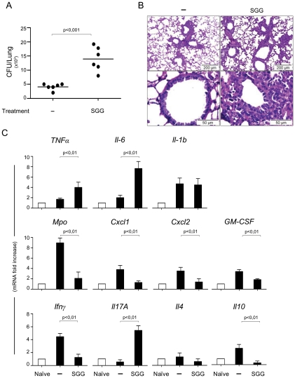
A new polysaccharide secreted by the human opportunistic fungal pathogen Aspergillus fumigatus has been characterized. Carbohydrate analysis using specific chemical degradations, mass spectrometry, ¹H and ¹³C nuclear magnetic resonance showed that this polysaccharide is a linear heterogeneous galactosaminogalactan composed of α1-4 linked galactose and α1-4 linked N-acetylgalactosamine residues where both monosacharides are randomly distributed and where the percentage of galactose per chain varied from 15 to 60%. This polysaccharide is antigenic and is recognized by a majority of the human population irrespectively of the occurrence of an Aspergillus infection. GalNAc oligosaccharides are an essential epitope of the galactosaminogalactan that explains the universal antibody reaction due to cross reactivity with other antigenic molecules containing GalNAc stretches such as the N-glycans of Campylobacter jejuni. The galactosaminogalactan has no protective effect during Aspergillus infections. Most importantly, the polysaccharide promotes fungal development in immunocompetent mice due to its immunosuppressive activity associated with disminished neutrophil infiltrates.


Conflict of interest statement

The authors have declared that no competing interests exist.

Figures

Figure 1
Figure 1. Detection of the galactosaminogalactan by immunofluorescence on resting, germinated conidia and on mycelium.
The specificity of the cell wall labelling was confirmed by the full inhibition of the labelling with the anti-GG MAb recognition seen after the MAb was incubated with GalNAc oligosaccharides obtained by HCl hydrolysis galactosaminogalactan (last panel on the right).
Figure 2
Figure 2. Analysis of periodate-oxidized galactosaminogalactan of A. fumigatus.
A, Gel permeation chromatography pattern of solubilised products on a HW40S column eluted with a 0.25% acetic acid solution. The three carbohydrate containing fractions (I-III) were identified by the refractometry index (RI). B, Composition of the oligosaccharides of fraction I purified on the HW40S gel filtration; composition was based on MALDI-TOF mass spectra (mass m/z  =  [M+Na]+); Th: threitol (from Galactose degradation); GalNAc: N-acetylgalactosamine.
Figure 3
Figure 3. Analysis of de-N-deacetylated and nitrous deaminated galactosaminogalactan of A. fumigatus.
A, Gel permeation chromatography pattern of solubilised products on a HW40S column eluted with a 0.25% acetic acid solution. The carbohydrate containing fractions were detected by the colorimetric phenol-sulphuric acid method. B, Composition of the oligosaccharides of fraction I purified on the HW40S gel filtration. The composition was based on MALDI-TOF mass spectra (mass m/z  =  [M+Na]+); AHT: 2,5-anhydrotalose (from GalNAc degradation); Gal: Galactose.
Figure 4
Figure 4. Vaccine potential of the urea soluble galactosaminogalactan (SGG) of A. fumigatus against invasive pulmonary aspergillosis.
A, C57BL/6 mice were injected with 2×10⁷ Aspergillus conidia 14 days or with CpG (10 nM) or CpG and SGG (5 µg) (CpG+SGG) 14, 7 and 3 days before the intranasal infection with 2 × 10⁷ live resting conidia. Naïve are uninfected mice and – are infected, untreated mice. Fungal growth is expressed as CFUs per lung and statistical significance is indicated by a p value <0.001. B, Bronchoalveolar cells were obtained by lung lavage and lung histology (PAS-staining) was done 3 days after infection. Note that SGG failed to ameliorate inflammatory pathology and even favoured fungal growth (insert) in the absence of neutrophil recruitment. C, Cytokines were determined by RT-PCR in lung homogenates 3 days after the infection. Results pooled from 2 experiments (6 animals/group). Photographs were taken using a high-resolution Microscopy Color Camera AxioCam. Bars indicate SEM and statistical significance is indicated by p values.
Figure 5
Figure 5. Impact of SGG on primary aspergillosis in intact mice.
A, C57BL/6 mice were first injected with SGG at day 3, 2 and 1 before conidial inhalation and infected on day 0 with 2×10⁷ Aspergillus conidia. Naïve are uninfected mice, – are infected, untreated mice and SGG are mice that have received SGG (250 mg/kg i.n. the day of the infection and on days 1, 2 and 3 post-infection). Fungal growth is expressed as CFUs per lung and statistical significance is indicated by a p value <0.001. Results pooled from 2 experiments (6 animals/group) with one example shown in panel A. B, Lung histology (PAS-staining) of mice treated as indicated, 3 days after infection showing signs of inflammatory pathology in the immunocompetent mice treated with SGG. C, Cytokines were determined by RT-PCR in lung homogenates 3 days after the infection. Note that SGG induced inflammatory cytokine gene expression, such as Tnfα, Il6, Il17a and Il4 genes, but suppressed Ifnγ and Il10 expression; the low level of Mpo gene expression is in agreement with the low counts of neutrophils in the lung of infected mice. Bars indicate SEM and statistical significance is indicated by p values.
Figure 6
Figure 6. SGG induces neutrophil apoptosis.
Whole-blood samples (500 µl) were incubated in 24-well tissue culture plates at 37°C for 20 h with 5% CO2 with PBS or SGG (1–20 µg/ml). Cycloheximide (CHX) (10 µg/ml) and GM-CSF (1000 pg/ml) were used as proapoptotic and antiapoptotic controls, respectively. Samples (100 µl) were incubated with APC-conjugated anti-CD15 and stained with FITC-conjugated annexin V and 7-AAD as described in Materials and Methods. Results are expressed as the percentage of total apoptotic cells (early and late apoptotic cells). Values are means ± SEM (n = 4). *Significantly different from sample incubated with PBS (p< 0.05). ° Significantly different from sample incubated with GM-CSF (p<0.05).
Figure 7
Figure 7. Binding of macrophage galactose-type lectin (MGL) to the galactosaminogalactan of A. fumigatus.
ELISA-inhibition of MGL-Fc interacting with α-GalNAc-conjugated polyacrylamide (GalNAc-PAA) by free GalNAc monosaccharides and a mixture of oligosaccharides exclusively composed of N-acetylgalactosamine with an average degree of polymerisation of 7.5 (G25-I). ELISA plates were coated with GalNAc-PAA (2 µg/ml). Plates were blocked with 1% BSA and the recombinant MGL-Fc chimera was added (0.5 µg/ml) for 2 h at room temperature in the presence of 0.125 and 0.25 mM of SGG hydrolysate fractions (G25-I). Binding was detected using a peroxidase-labeled anti-human IgG-Fc antibody.

References

    1. Morgan J, Wannemuehler KA, Marr KA, Hadley S, Kontoyiannis DP, et al. Incidence of invasive aspergillosis following hematopoietic stem cell and solid organ transplantation: interim results of a prospective multicenter surveillance program. Med Mycol. 2005;43(Suppl 1):S49–58. - PubMed
    1. Singh N, Paterson DL. Aspergillus infections in transplant recipients. Clin Microbiol Rev. 2005;18:44–69. - PMC - PubMed
    1. Brakhage AA, Bruns S, Thywissen A, Zipfel PF, Behnsen J. Interaction of phagocytes with filamentous fungi. Curr Opin Microbiol. 2010;13:409–415. - PubMed
    1. Bozza S, Clavaud C, Giovannini G, Fontaine T, Beauvais A, et al. Immune sensing of Aspergillus fumigatus proteins, glycolipids, and polysaccharides and the impact on Th immunity and vaccination. J Immunol. 2009;183:2407–2414. - PubMed
    1. Mouyna I, Fontaine T. Cell wall of Aspergillus fumigatus: a dynamic structure. In: Latgé JP, Steinbach WJ, editors. Washington, D.C: American Society for Microbiology; 2009. pp. 169–183.

Publication types

MeSH terms