. 2011 Nov;7(11):e1002381.
doi: 10.1371/journal.ppat.1002381. Epub 2011 Nov 10.

Cross-reactive T cells are involved in rapid clearance of 2009 pandemic H1N1 influenza virus in nonhuman primates

Affiliations

Cross-reactive T cells are involved in rapid clearance of 2009 pandemic H1N1 influenza virus in nonhuman primates

Jason T Weinfurter et al. PLoS Pathog. 2011 Nov.

Abstract

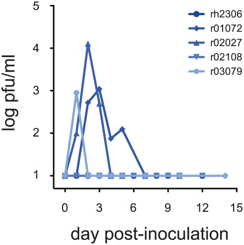
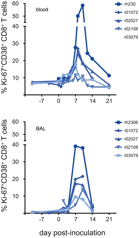
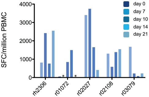
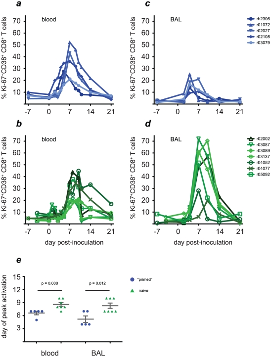
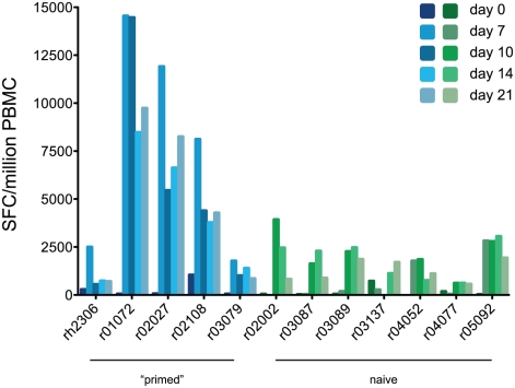
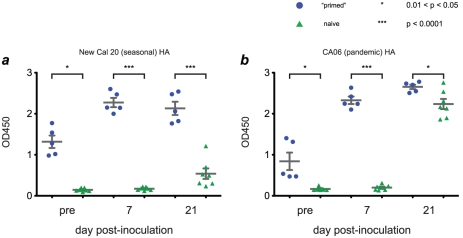
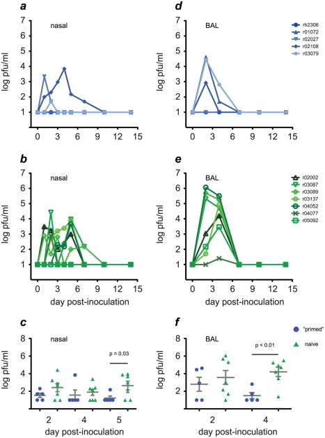
In mouse models of influenza, T cells can confer broad protection against multiple viral subtypes when antibodies raised against a single subtype fail to do so. However, the role of T cells in protecting humans against influenza remains unclear. Here we employ a translational nonhuman primate model to show that cross-reactive T cell responses play an important role in early clearance of infection with 2009 pandemic H1N1 influenza virus (H1N1pdm). To "prime" cellular immunity, we first infected 5 rhesus macaques with a seasonal human H1N1 isolate. These animals made detectable cellular and antibody responses against the seasonal H1N1 isolate but had no neutralizing antibodies against H1N1pdm. Four months later, we challenged the 5 "primed" animals and 7 naive controls with H1N1pdm. In naive animals, CD8+ T cells with an activated phenotype (Ki-67+ CD38+) appeared in blood and lung 5-7 days post inoculation (p.i.) with H1N1pdm and reached peak magnitude 7-10 days p.i. In contrast, activated T cells were recruited to the lung as early as 2 days p.i. in "primed" animals, and reached peak frequencies in blood and lung 4-7 days p.i. Interferon (IFN)-γ Elispot and intracellular cytokine staining assays showed that the virus-specific response peaked earlier and reached a higher magnitude in "primed" animals than in naive animals. This response involved both CD4+ and CD8+ T cells. Strikingly, "primed" animals cleared H1N1pdm infection significantly earlier from the upper and lower respiratory tract than the naive animals did, and before the appearance of H1N1pdm-specific neutralizing antibodies. Together, our results suggest that cross-reactive T cell responses can mediate early clearance of an antigenically novel influenza virus in primates. Vaccines capable of inducing such cross-reactive T cells may help protect humans against severe disease caused by newly emerging pandemic influenza viruses.


Conflict of interest statement

The authors have declared that no competing interests exist.

Figures

Figure 1
Figure 1. A seasonal human influenza virus replicates in rhesus macaques.
Five rhesus macaques were inoculated with 9 million plaque-forming units (pfu) influenza virus A/Kawasaki/173/2001 (K173, H1N1) by a combination of routes as described in the text. Animals had no detectable antibodies against K173 at the time of inoculation. Virus replication in nasal secretions was monitored using standard plaque assays. Virus titer is expressed as pfu/ml nasal swab fluid.
Figure 2
Figure 2. Activated CD8+ T cells appear in blood and lung within 7 days of inoculation with seasonal influenza virus K173.
We used a flow cytometric assay to enumerate CD8+ T cells with an activated phenotype before and after K173 infection as described in the text. Data plotted are the frequency of CD3+ CD8+ lymphocytes that express both Ki-67 and CD38. Data are not available from animal r01072 at day 21 post-inoculation due to technical problems.
Figure 3
Figure 3. Macaques mount virus-specific T cell responses after K173 infection.
We used an IFN-γ Elispot assay to enumerate influenza virus-specific T cells among the peripheral blood mononuclear cells (PBMC) of infected macaques. PBMC were stimulated with 19 pools of overlapping synthetic peptides that collectively represented the influenza virus proteome, as described in the text. After subtracting background signal from the results for each pool, the frequency of cells responding to each pool were summed to give the total virus-specific response magnitude. Results are expressed as the number of spot-forming cells (SFC) per million PBMC. To be considered positive, the frequency of peptide-specific SFC/million PBMC must be at least 3 times larger than the background (no peptide) value. Asterisks indicate that data are not available from animal r01072 from days 7 or 21 post-inoculation due to technical problems.
Figure 4
Figure 4. Activated CD8+ T cell populations increase rapidly in the blood and lungs of “primed” animals after challenge with H1N1pdm.
Four months after infection with the K173 seasonal isolate, we challenged the 5 previously infected macaques (blue traces, solid symbols) and 7 naive control animals (green traces, open symbols) with 9 million pfu of H1N1pdm isolate A/California/04/2009 (CA04) and monitored the kinetics of CD8+ T cell activation. a and b, CD8+ T cell activation kinetics in blood. c and d, CD8+ T cell activation kinetics in lung, assessed using cells recovered from bronchoalveolar lavage (BAL). e, Peak levels of activation are reached significantly earlier in both compartments in “primed” animals than in naive animals.
Figure 5
Figure 5. “Primed” animals mount stronger virus-specific T cell responses than naive animals after H1N1pdm challenge.
T cell responses against the viral proteome were measured using IFN-γ Elispot as described in Fig. 3. Blue bars, “primed” animals; Green bars, naive animals.
Figure 6
Figure 6. Infection with seasonal influenza viruses induces variable levels of antibody capable of binding H1N1pdm antigens.
We used purified hemagglutinin (HA) proteins to capture antibodies from macaque serum. Antibodies were detected using an anti-IgG antibody coupled to horseradish peroxidase, with enzymatic activity quantified using a colorimetric assay. Results are expressed in terms of sample absorbance at 450 nm. Antigens used in each assay were as follows: a, Purified HA protein from A/New Caledonia/20/1999, a close antigenic match to K173. b, Purified HA from A/California/06/2009, a close antigenic match to CA04. Assays using lysates of K173 or CA04 viruses gave similar results (data not shown).
Figure 7
Figure 7. “Primed” animals clear H1N1pdm infection more rapidly than naive animals.
We used plaque assays to monitor replication of H1N1pdm in the upper respiratory tract (nasal secretions) and lower respiratory tract (BAL) in “primed” (blue traces, solid symbols) and naive animals (green traces, open symbols). a and b, kinetics of H1N1pdm replication in nasal secretions. c, By day 5 post-inoculation virus titers in nasal secretions were significantly lower in “primed” than naive animals. d and e, H1N1pdm replication kinetics in the lung. f, By day 4 post-infection virus titers in lung were significantly lower in “primed” than naive animals.

References

    1. Fraser C, Donnelly CA, Cauchemez S, Hanage WP, Van Kerkhove MD, et al. Pandemic potential of a strain of influenza A (H1N1): early findings. Science. 2009;324:1557–1561. - PMC - PubMed
    1. Neumann G, Noda T, Kawaoka Y. Emergence and pandemic potential of swine-origin H1N1 influenza virus. Nature. 2009;459:931–939. - PMC - PubMed
    1. US Centers for Disease Control and Prevention. Serum cross-reactive antibody response to a novel influenza A (H1N1) virus after vaccination with seasonal influenza vaccine. MMWR Morb Mortal Wkly Rep. 2009;58:521–524. - PubMed
    1. Hancock K, Veguilla V, Lu X, Zhong W, Butler EN, et al. Cross-reactive antibody responses to the 2009 pandemic H1N1 influenza virus. N Engl J Med. 2009;361:1945–1952. - PubMed
    1. Itoh Y, Shinya K, Kiso M, Watanabe T, Sakoda Y, et al. In vitro and in vivo characterization of new swine-origin H1N1 influenza viruses. Nature. 2009;460:1021–1025. - PMC - PubMed

Publication types

MeSH terms