

Identifying a network of brain regions involved in aversion-related processing: a cross-species translational investigation
- PMID: 22102836
- PMCID: PMC3215229
- DOI: 10.3389/fnint.2011.00049
Identifying a network of brain regions involved in aversion-related processing: a cross-species translational investigation
Abstract
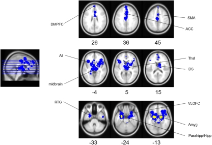
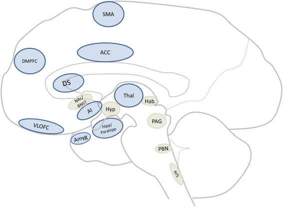
The ability to detect and respond appropriately to aversive stimuli is essential for all organisms, from fruit flies to humans. This suggests the existence of a core neural network which mediates aversion-related processing. Human imaging studies on aversion have highlighted the involvement of various cortical regions, such as the prefrontal cortex, while animal studies have focused largely on subcortical regions like the periaqueductal gray and hypothalamus. However, whether and how these regions form a core neural network of aversion remains unclear. To help determine this, a translational cross-species investigation in humans (i.e., meta-analysis) and other animals (i.e., systematic review of functional neuroanatomy) was performed. Our results highlighted the recruitment of the anterior cingulate cortex, the anterior insula, and the amygdala as well as other subcortical (e.g., thalamus, midbrain) and cortical (e.g., orbitofrontal) regions in both animals and humans. Importantly, involvement of these regions remained independent of sensory modality. This study provides evidence for a core neural network mediating aversion in both animals and humans. This not only contributes to our understanding of the trans-species neural correlates of aversion but may also carry important implications for psychiatric disorders where abnormal aversive behavior can often be observed.
Keywords: animal models; aversion; imaging; meta-analysis; translational.
Figures
References
-
- Albrechet-Souza L., Borelli K. G., Carvalho M. C., Brandao M. L. (2009). The anterior cingulate cortex is a target structure for the anxiolytic-like effects of benzodiazepines assessed by repeated exposure to the elevated plus maze and Fos immunoreactivity. Neuroscience 164, 387–39710.1016/j.neuroscience.2009.08.038 - DOI - PubMed
References Used in Meta-Analysis
-
- Botvinick M., Jha A. P., Bylsma L. M., Fabian S. A., Solomon P. E., Prkachin K. M. (2005). Viewing facial expressions of pain engages cortical areas involved in the direct experience of pain. Neuroimage 25, 312–319 - PubMed
-
- Buchel C., Bornhovd K., Quante M., Glauche V., Bromm B., Weiller C. (2002). Dissociable neural responses related to pain intensity, stimulus intensity, and stimulus awareness within the anterior cingulate cortex: a parametric single-trial laser functional magnetic resonance imaging study. J. Neurosci. 22, 970–976 - PMC - PubMed
LinkOut - more resources
Full Text Sources