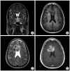
Supratentorial clear cell ependymoma mimicking oligodendroglioma : case report and review of the literature
- PMID: 22102956
- PMCID: PMC3218185
- DOI: 10.3340/jkns.2011.50.3.240
Supratentorial clear cell ependymoma mimicking oligodendroglioma : case report and review of the literature
Abstract
Clear cell ependymomas (CCEs) are rare variants of ependymomas. Tumors show anaplastic histological features and behave as an aggressive manner. CCEs have a predilection for extraneural metastases and early recurrence, and they demonstrate characteristic radiographic features. These tumors should be radiologically and pathologically differentiated from oligodendrogliomas. On microscopic examination, CCEs are composed of sheets of cells and resemble oligodendroglioma. However, upon closer examination, the nature of CCEs can be detected earlier, resulting in prompt treatment of the tumor. Although we report only one case, we emphasize the importance of early diagnosis and treatment. Future description of more cases of these rare cancers is necessary to aid in their diagnosis and treatment.
Keywords: Clear cell; Ependymoma; Histology; Oligodendroglioma; Prognosis.
Figures


Similar articles
-
Clear cell ependymoma: a mimic of oligodendroglioma: clinicopathologic and ultrastructural considerations.Am J Surg Pathol. 1997 Jul;21(7):820-6. doi: 10.1097/00000478-199707000-00010. Am J Surg Pathol. 1997. PMID: 9236838 Review.
-
Clear cell ependymoma: a mimicker of oligodendroglioma--report of three cases.Neuropathology. 2008 Aug;28(4):366-71. doi: 10.1111/j.1440-1789.2008.00895.x. Epub 2008 Feb 1. Neuropathology. 2008. PMID: 18248576
-
Clear cell ependymoma: a clinicopathologic and radiographic analysis of 10 patients.Cancer. 2003 Nov 15;98(10):2232-44. doi: 10.1002/cncr.11783. Cancer. 2003. PMID: 14601094
-
Value and limits of immunohistochemistry in differential diagnosis of clear cell primary brain tumors.Acta Neuropathol. 2004 Jul;108(1):24-30. doi: 10.1007/s00401-004-0856-9. Epub 2004 Apr 23. Acta Neuropathol. 2004. PMID: 15108012
-
Supratentorial cortical ependymoma: case series and review of the literature.Neuropathology. 2014 Jun;34(3):243-52. doi: 10.1111/neup.12087. Epub 2013 Dec 20. Neuropathology. 2014. PMID: 24354554 Review.
Cited by
-
Prognostic Implications of Histological Clear Cells in High-Grade Intracranial Ependymal Tumors: A Retrospective Analysis from a Tertiary Care Hospital in Pakistan.Asian J Neurosurg. 2018 Apr-Jun;13(2):307-313. doi: 10.4103/ajns.AJNS_280_16. Asian J Neurosurg. 2018. PMID: 29682026 Free PMC article.
-
Ependymal tumors with oligodendroglioma like clear cells: Experience from a tertiary care hospital in Pakistan.Surg Neurol Int. 2015 Nov 16;6(Suppl 23):S583-9. doi: 10.4103/2152-7806.169545. eCollection 2015. Surg Neurol Int. 2015. PMID: 26664928 Free PMC article.
-
Supra-sellar clear cell ependymoma in a 2-year-old female: A case report.Int J Surg Case Rep. 2025 Jul;132:111446. doi: 10.1016/j.ijscr.2025.111446. Epub 2025 May 14. Int J Surg Case Rep. 2025. PMID: 40398199 Free PMC article.
-
Cerebellar ependymoma with overlapping features of clear-cell and tanycytic variants mimicking hemangioblastoma: a case report and literature review.Diagn Pathol. 2017 Mar 20;12(1):28. doi: 10.1186/s13000-017-0619-2. Diagn Pathol. 2017. PMID: 28320419 Free PMC article. Review.
References
-
- Amatya VJ, Takeshima Y, Kaneko M, Nakano T, Yamaguchi S, Sugiyama K, et al. Case of clear cell ependymoma of medulla oblongata : clinicopathological and immunohistochemical study with literature review. Pathol Int. 2003;53:297–302. - PubMed
-
- Fokes EC, Jr, Earle KM. Ependymomas : clinical and pathological aspects. J Neurosurg. 1969;30:585–594. - PubMed
-
- Fouladi M, Helton K, Dalton J, Gilger E, Gajjar A, Merchant T, et al. Clear cell ependymoma : a clinicopathologic and radiographic analysis of 10 patients. Cancer. 2003;98:2232–2244. - PubMed
-
- Gilbert MR, Ruda R, Soffietti R. Ependymomas in adults. Curr Neurol Neurosci Rep. 2010;10:240–247. - PubMed
-
- Jain D, Sharma MC, Arora R, Sarkar C, Suri V. Clear cell ependymoma : a mimicker of oligodendroglioma-report of three cases. Neuropathology. 2008;28:366–371. - PubMed
Publication types
LinkOut - more resources
Full Text Sources

