. 2011 Nov;34(10):1584-94.
doi: 10.1111/j.1460-9568.2011.07896.x.

Differential gene expression in migratory streams of cortical interneurons

Affiliations
Free PMC article

Differential gene expression in migratory streams of cortical interneurons

Mary Antypa et al. Eur J Neurosci. 2011 Nov.
Free PMC article

Abstract

Cortical interneurons originate in the ganglionic eminences of the subpallium and migrate into the cortex in well-defined tangential streams. At the start of corticogenesis, two streams of migrating neurons are evident: a superficial one at the level of the preplate (PPL), and a deeper one at the level of the intermediate zone (IZ). Currently, little is known about the signalling mechanisms that regulate interneuron migration, and almost nothing is known about the molecules that may be involved in their choice of migratory stream. Here, we performed a microarray analysis, comparing the changes in gene expression between cells migrating in the PPL and those migrating in the IZ at embryonic day 13.5. This analysis identified genes, many of them novel, that were upregulated in one of the two streams. Moreover, polymerase chain reaction, in situ hybridization experiments and immunohistochemistry showed the expression of these genes in interneurons migrating within the PPL or IZ, suggesting that they play a role in their migration and choice of stream.


Figures

Figure 1
Figure 1
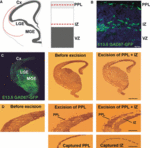
Tangential migration of interneurons into the cerebral cortex. (A) Schematic diagrams depicting the streams of migrating interneurons at E13.5. Red lines indicate the laminar positions of the PPL and IZ streams. (B) Coronal section through the cortex of an E13.5 GAD67–GFP transgenic mouse showing abundant migrating cells in the PPL and IZ streams. (C and D) Intact section through the forebrain of one hemisphere of an E13.5 GAD67–GFP transgenic mouse and after excision and capture of the PPL and IZ with a laser-capture microscope. Scale bars: (A) 100 μm; (C and D) 500 μm. Cx, cerebral cortex; LGE, lateral ganglionic eminence; VZ, ventricular zone.
Figure 2
Figure 2
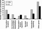
Numbers of genes upregulated in the PPL (grey bars) and IZ (black bars) migratory streams at E13.5. Genes were classified into the categories listed according to their molecular function.
Figure 3
Figure 3
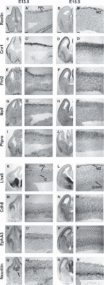
Expression of receptor genes in the interneuron migratory streams in the developing forebrain as seen by in situ hybridization at E13.5 and E15.5. A higher-magnification image of the cortex is shown beside each low-magnification panel of the forebrain. The upper panels show the expression of receptor genes in the PPL at E13.5 and MZ at E15.5. The lower panels show the expression of receptor genes predominantly in the IZ at E13.5 and E15.5. (A–B′) The expression of Reelin was used as an internal control, as it is known that it is expressed exclusively in cells (presumptive CR cells) in the PPL at E13.5 and in the MZ at E15.5. (C–J′) Expression of receptor genes Cnr1 (C–D′), Flrt2 (E–F′), Nelf (G–H′) and Ptpro (I–J) was localized predominantly within the PPL at E13.5 and within the MZ at E15.5. (K–L′) The interneuron marker Lhx6 was also used as an internal control, as it is known to be expressed in both the PPL and IZ at E13.5 (K–K′), and more widely, but predominantly in the MZ and IZ/SVZ, at E15.5 (L–L′). (M–R′) Expression of the receptor genes Cdh8 (M–N′), EphA3 (O–P′) and Neuritin (Q–R′) within the IZ at E13.5 and within the IZ/SVZ at E15.5. Scale bar in A–B′: 200 μm.
Figure 4
Figure 4
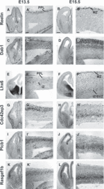
Expression of cell signalling genes in the interneuron migratory streams in the developing forebrain as seen by in situ hybridization at E13.5 and E15.5. A higher-magnification image of the cortex is shown beside each low-magnification panel of the forebrain. The upper panels show the expression of receptor genes in the PPL at E13.5 and in the MZ at E15.5. The lower panels show the expression of receptor genes predominantly in the IZ at E13.5 and E15.5. (A–B′) The expression of Reelin was used as an internal control, as it is known that it is expressed exclusively in cells (presumptive CR cells) in the PPL at E13.5 and in the MZ at E15.5. (C–D′) Expression of Dab1 is observed only within the PPL at E13.5 and in the MZ and SP (after the splitting of the CP) at E15.5. (E–F′) The interneuron marker Lhx6 was also used as an internal control, as it is known to be expressed in both the PPL and IZ at E13.5 (E–E′), and more widely, but predominantly in the MZ and IZ/SVZ, at E15.5 (F–F′). (G–L′) Expression of the cell signalling genes Cdc42ep3 (G–H′), Plcb1 (I–J′) and Rasgef1b (K–L′) was observed predominantly within the IZ at E13.5, and within the IZ/SVZ at E15.5. Scale bar in A–B′: 200 μm.
Figure 5
Figure 5
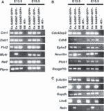
Agarose gel electophoresis of qPCR products. qPCR was performed on cortex-derived and GE-derived GAD67–GFP-positive and GAD67–GFP-negative cells at E13.5 and E15.5. The genes examined are listed next to the gel bands. (A) Genes shown to be upregulated in the cortical PPL/MZ. Nearly all genes were expressed in the three samples at both ages. The only exception was Mc4r, which was found to be expressed only in GAD67–GFP-negative cells (presumptive pyramidal neuron progenitors) in the cortex at E13.5. (B) Genes shown to be upregulated in the IZ. (C) Control genes. Cx, cortex; -RT, without reverse transcriptase (negative control).
Figure 6
Figure 6
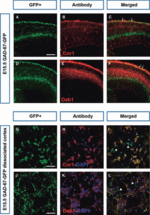
Expression of Cnr1 and Dab1 in interneurons. (A–F) Coronal sections from GAD67–GFP-positive mice at E15.5 were immunostained with anti-Cnr1 (B) and anti-Dab1 (E) (red). Co-localization (yellow; arrows) of Cnr1 and Dab1 with GFP is evident in some neurons in the MZ in the merged panels (C and F). (G–L) Dissociated cortical cell cultures prepared from E15.5 GAD67–GFP-positive cortices were immunostained with anti-Cnr1 (H) and anti-Dab1 (K). GFP single-positive cells are indicated by arrowheads, and Cnr1/GFP or Dab1/GFP double-positive cells are indicated by arrows in the merged panels (I and L). Scale bars: (A) 200 μm; (G) 15 μm.

Similar articles

Cited by

References

    1. Alifragis P, Liapi A, Parnavelas JG. Lhx6 regulates the migration of cortical interneurons from the ventral telencephalon but does not specify their GABA phenotype. J. Neurosci. 2004;24:5643–5648. - PMC - PubMed
    1. Anderson SA, Marín O, Horn C, Jennings K, Rubenstein JL. Distinct cortical migrations from the medial and lateral ganglionic eminences. Development. 2001;128:353–363. - PubMed
    1. Andrews W, Liapi A, Plachez C, Camurri L, Zhang J, Mori S, Murakami F, Parnavelas JG, Sundaresan V, Richards LJ. Robo1 regulates the development of major axon tracts and interneuron migration in the forebrain. Development. 2006;133:2243–2252. - PubMed
    1. Andrews WD, Barber M, Parnavelas JG. Slit–Robo interactions during cortical development. J. Anat. 2007;211:188–198. - PMC - PubMed
    1. Andrews W, Barber M, Hernadez-Miranda LR, Xian J, Rakic S, Sundaresan V, Rabbitts TH, Pannell R, Rabbitts P, Thompson H, Erskine L, Murakami F, Parnavelas JG. The role of Slit–Robo signaling in the generation, migration and morphological differentiation of cortical interneurons. Dev. Biol. 2008;313:648–658. - PubMed

Publication types

MeSH terms

Substances