Review
. 2011 Nov;34(10):1663-76.
doi: 10.1111/j.1460-9568.2011.07892.x.

What can we get from 'barrels': the rodent barrel cortex as a model for studying the establishment of neural circuits

Affiliations
Review

What can we get from 'barrels': the rodent barrel cortex as a model for studying the establishment of neural circuits

Chia-Shan Wu et al. Eur J Neurosci. 2011 Nov.

Abstract

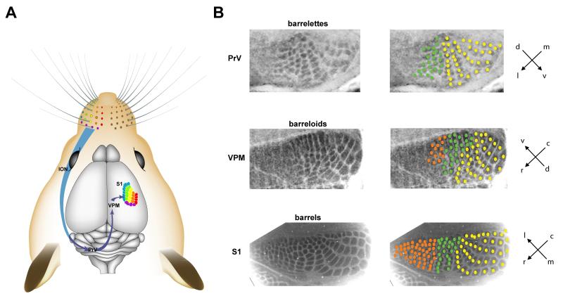
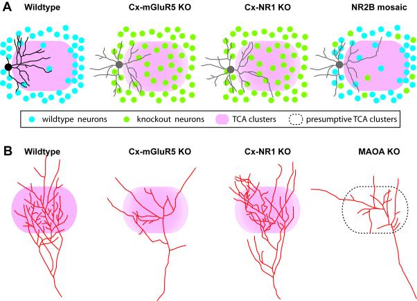
Sensory inputs triggered by external stimuli are projected into discrete arrays of neuronal modules in the primary sensory cortex. This whisker-to-barrel pathway has gained in popularity as a model system for studying the development of cortical circuits and sensory processing because its clear patterns facilitate the identification of genetically modified mice with whisker map deficits and make possible coordinated in vitro and in vivo electrophysiological studies. Numerous whisker map determinants have been identified in the past two decades. In this review, we summarize what have we learned from the detailed studies conducted in various mutant mice with cortical whisker map deficits. We will specifically focus on the anatomical and functional establishment of the somatosensory thalamocortical circuits.


Figures

Figure 1
Figure 1
The lemniscal whisker-to-barrel pathway in the mouse brain. (A) The whiskers on the snout are innervated by the infraorbital branch of the maxillary nerve (ION), which transmit sensory information to the rostral principal nucleus (PrV) in the brainstem. Trigeminothalamic axons from the PrV project to the ventral posteromedial nucleus (VPM) in the thalamus in the contralateral hemisphere. Thalamocortical axons from the VPM project to the primary somatosensory cortex (S1). The five rows of whiskers and the straddle whiskers are color-coded. (B) Representative images of CO-stained coronal sections through PrV of brain stem (barrelettes), VPM of thalamus (barreloids), and tangential sections through cortical layer IV (barrels). CO patches corresponding to rostroventral whiskers are marked with orange and green circles, and caudodorsal whiskers with yellow circles. Drawing in A is modified from an Adobe Illustrator file generously provided by Dr. Knott (Knott et al., 2002).
Figure 2
Figure 2
Timeline for development of the mouse cortical whisker map.
Figure 3
Figure 3
Schematic diagram showing examples of reconstructed layer IV spiny stellate neurons, and single thalamocortical axons in wildtype, Cx-mGluR5 KO, Cx-NR1 KO and NR2B mosaic mutant mice. (A) In wildtype mice, layer IV spiny stellate neurons show polarized dendritic morphology with the majority of their dendrites projecting toward the barrel hollow (pink). For simplicity, only spiny stellate neurons are depicted here. In both Cx-mGluR5 and Cx-NR1 KO mice, layer IV neurons are evenly distributed in the barrel field, and have symmetric dendritic morphology (non-polarized pattern, depicted in grey). In NR2B mosaic KO mice, mutant layer IV neurons located in the barrel walls show non-polarized dendritic pattern. (B) Examples of reconstructed single TCAs. In wildtype mice, the axon arbor in layer IV develops from a single axon, forming numerous axon collaterals, with a dominant orientation of the branches toward the barrel center, individual TCAs form highly branched and densely clustered arbors corresponding to the mapped facial whisker. In Cx-mGluR5 KO mice and MAOA KO mice, the complexity of TCA arbors is much reduced, and collaterals grow in divergent directions instead of forming a narrow cluster. In Cx-NR1 KO mice, TCAs have exuberant branches. Drawings were based on data shown in Datwani et al. (2002), Rebsam et al. (2002), Lee et al. (2005), Espinosa et al. (2009), and Ballester-Rosado et al. (2010).

Similar articles

Cited by

References

    1. Abdel-Majid RM, Leong WL, Schalkwyk LC, Smallman DS, Wong ST, Storm DR, Fine A, Dobson MJ, Guernsey DL, Neumann PE. Loss of adenylyl cyclase I activity disrupts patterning of mouse somatosensory cortex. Nat Genet. 1998;19:289–291. - PubMed
    1. Agmon A, Connors BW. Thalamocortical responses of mouse somatosensory (barrel) cortex in vitro. Neuroscience. 1991;41:365–379. - PubMed
    1. Agmon A, O’Dowd DK. NMDA receptor-mediated currents are prominent in the thalamocortical synaptic response before maturation of inhibition. J Neurophysiol. 1992;68:345–349. - PubMed
    1. Agmon A, Yang LT, Jones EG, O’Dowd DK. Topological precision in the thalamic projection to neonatal mouse barrel cortex. J Neurosci. 1995;15:549–561. - PMC - PubMed
    1. Alagarsamy S, Rouse ST, Junge C, Hubert GW, Gutman D, Smith Y, Conn PJ. NMDA-induced phosphorylation and regulation of mGluR5. Pharmacol Biochem Behav. 2002;73:299–306. - PubMed

Publication types