. 2011 Dec 21;133(50):20357-68.
doi: 10.1021/ja207407n. Epub 2011 Nov 21.

Structure of (5'S)-8,5'-cyclo-2'-deoxyguanosine in DNA

Affiliations

Structure of (5'S)-8,5'-cyclo-2'-deoxyguanosine in DNA

Hai Huang et al. J Am Chem Soc. .

Abstract

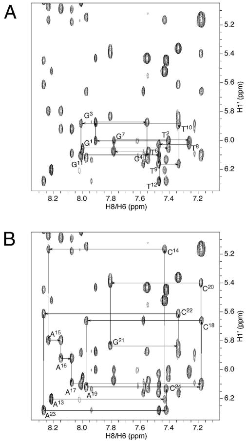
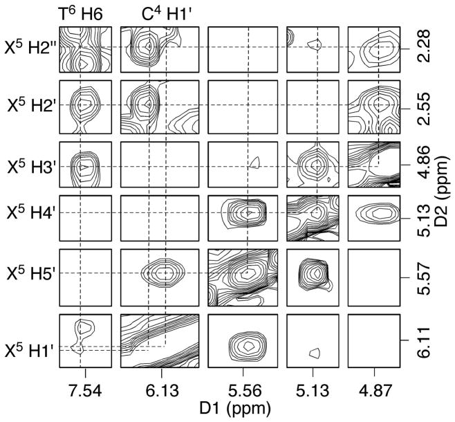
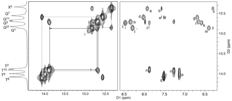
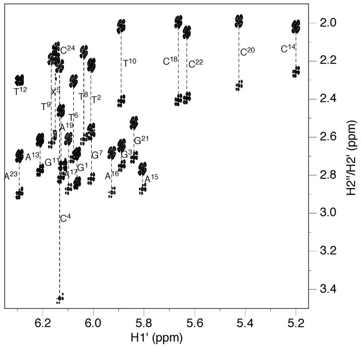
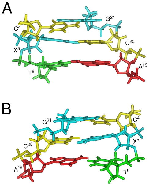
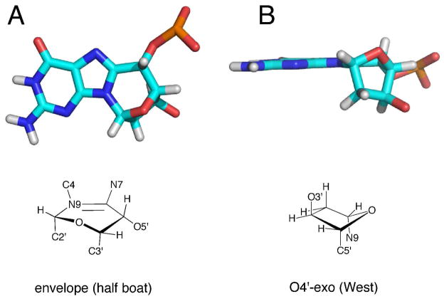
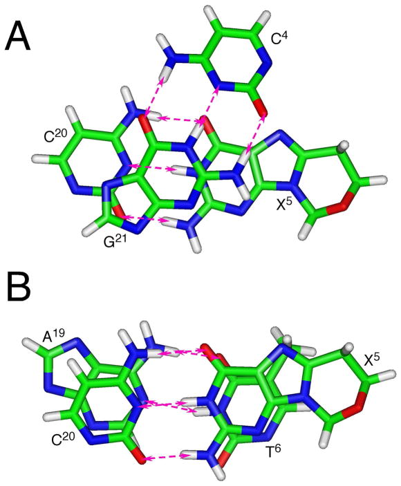
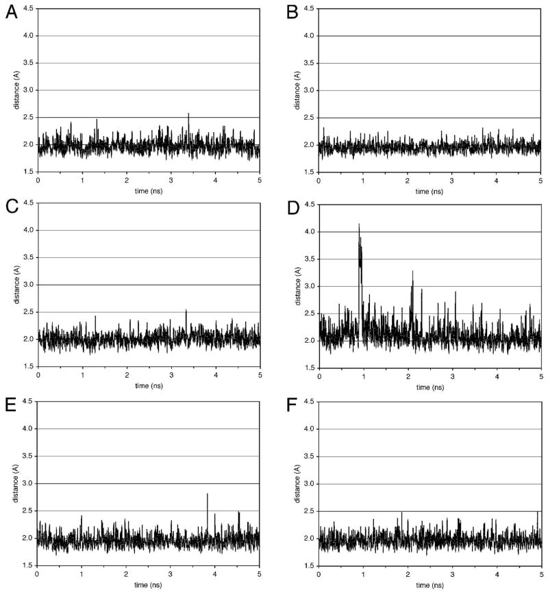
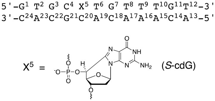
Diastereomeric 8,5'-cyclopurine 2'-deoxynucleosides, containing a covalent bond between the deoxyribose and the purine base, represent an important class of DNA damage induced by ionizing radiation. The 8,5'-cyclo-2'-deoxyguanosine lesion (cdG) has been recently reported to be a strong block of replication and highly mutagenic in Escherichia coli. The 8,5'-cyclopurine-2'-deoxyriboses are suspected to play a role in the etiology of neurodegeneration in xeroderma pigmentosum patients. These lesions cannot be repaired by base excision repair, but they are substrates for nucleotide excision repair. The structure of an oligodeoxynucleotide duplex containing a site-specific S-cdG lesion placed opposite dC in the complementary strand was obtained by molecular dynamics calculations restrained by distance and dihedral angle restraints obtained from NMR spectroscopy. The S-cdG deoxyribose exhibited the O4'-exo (west) pseudorotation. Significant perturbations were observed for the β, γ, and χ torsion angles of the S-cdG nucleoside. Watson-Crick base pairing was conserved at the S-cdG·dC pair. However, the O4'-exo pseudorotation of the S-cdG deoxyribose perturbed the helical twist and base pair stacking at the lesion site and the 5'-neighbor dC·dG base pair. Thermodynamic destabilization of the duplex measured by UV melting experiments correlated with base stacking and structural perturbations involving the modified S-cdG·dC and 3'- neighbor dT·dA base pairs. These perturbations may be responsible for both the genotoxicity of this lesion and its ability to be recognized by nucleotide excision repair.


Figures

Figure 1
Figure 1
NOE connectivity of base H8/H6 protons with deoxyribose H1′ protons of the S-cdG containing duplex. A. Modified strand. B. Complementary strand.
Figure 2
Figure 2
Tile plot derived from a NOESY spectrum obtained at a mixing time of 60 ms showing the assignment of S-cdG non-exchangeable protons.
Figure 3
Figure 3
Assignment of the base imino and amino protons based on the NOE connectivity. NOE interactions of the imino protons with the opposite base arising from Watson-Crick base pairing are labeled as: (a) X5 N1H → C20 N4H2, (b) X5 N1H → A19 H2, (c) X5 N1H → C20 N4H1, (d) G7 N1H → C18 N4H2, (e) G7 N1H → A19 H2, (f) G7 N1H → A17 H2, (g) G7 N1H → C18 N4H1, (h) G11 N1H → C14 N4H2, (i) G11 N1H → C14 N4H1, (j) G3 N1H → C22 N4H2, (k) G3 N1H → C22 N4H1, (l) G21 N1H → C4 N4H2, (m) G21 N1H → C4 N4H1, (n) T2 N3H → A23 H2, (o) T10 N3H → A15 H2, (p) T9 N3H → A16 H2, and (q) T8 N3H → A17 H2.
Figure 4
Figure 4
An expansion of the ECOSY spectrum used for the measurement of 3JH1′-H2′ and 3JH1′-H2″ coupling constants. Except for X5, G11, and T12, all H2″ protons exhibited greater chemical shifts than H2′ protons. The geminal H2′ and H2″ protons of G11 and T12 were not resolved, and X5 H2″ was upfield from H2′.
Figure 5
Figure 5
31P NMR of the S-cdG containing duplex compared with the corresponding unmodified duplex. A. Unmodified duplex. B. S-cdG containing duplex.
Figure 6
Figure 6
Proton chemical shift perturbations of the S-cdG containing duplex compared with the unmodified duplex. A. Base protons of the S-cdG-modified strand. B. Deoxyribose protons of the S-cdG-modified strand. C. Base protons of the complementary strand. D. Deoxyribose protons of the complementary strand.
Figure 7
Figure 7
1H NMR of the S-cdG containing duplex compared with the corresponding unmodified duplex at different temperatures. A. Unmodified duplex. B. S-cdG containing duplex.
Figure 8
Figure 8
Expanded views of the refined structure of the S-cdG containing duplex at the lesion site. A. View from the major groove. B. View from minor groove.
Figure 9
Figure 9
Ring conformations of the S-cdG in the refined structure. A. Six-member ring C8-N9-C1′-O4′-C4′-C5′. B. 2′-deoxyribose.
Figure 10
Figure 10
Base pairing and base stacking of the refined structure of the S-cdG containing duplex at the lesion site. The pink arrows indicate anticipated hydrogen bonding interactions. A. the C4•G21 and X5•C20 base pairs. B. The X5•C20 and T6•A19 base pairs.
Figure 11
Figure 11
Distances of guanine N1H → cytosine N3 and the thymine N3H → adenine N1 of some base pairs in the trajectories of the molecular dynamics simulations conducted in explicit solvent at 300 K. A. T2•A23 base pair. B. C4•G21 base pair. C. X5•C20 base pair. D. T6•A19 base pair. E. T8•A17 base pair. F. T9•A16 base pair.
Scheme 1
Scheme 1
Numbering scheme of the oligodeoxynucleotide duplex containing the (5′S)-8,5′-cyclo-2′-deoxyguanosine (S-cdG) 5′-nucleotide.

Similar articles

Cited by

References

    1. Cooke MS, Evans MD, Dizdaroglu M, Lunec J. Faseb J. 2003;17:1195–1214. - PubMed
    1. Dedon PC. Chem Res Toxicol. 2008;21:206–219. - PubMed
    1. Keck K. Z Naturforsch B. 1968;23:1034–1043. - PubMed
    1. Jaruga P, Dizdaroglu M. DNA Repair (Amst) 2008;7:1413–1425. - PubMed
    1. Dizdaroglu M. Biochem J. 1986;238:247–254. - PMC - PubMed

Publication types