. 2012 Feb 17;7(2):378-86.
doi: 10.1021/cb200352q. Epub 2011 Dec 15.

Characterization of molecular interactions between ACP and halogenase domains in the Curacin A polyketide synthase

Affiliations

Characterization of molecular interactions between ACP and halogenase domains in the Curacin A polyketide synthase

Alena Busche et al. ACS Chem Biol. .

Abstract

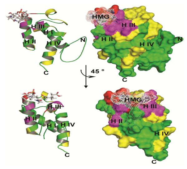
Polyketide synthases (PKSs) and non-ribosomal peptide synthetases (NRPSs) are large multidomain proteins present in microorganisms that produce bioactive compounds. Curacin A is such a bioactive compound with potent anti-proliferative activity. During its biosynthesis the growing substrate is bound covalently to an acyl carrier protein (ACP) that is able to access catalytic sites of neighboring domains for chain elongation and modification. While ACP domains usually occur as monomers, the curacin A cluster codes for a triplet ACP (ACP(I)-ACP(II)-ACP(III)) within the CurA PKS module. We have determined the structure of the isolated holo-ACP(I) and show that the ACPs are independent of each other within this tridomain system. In addition, we have determined the structure of the 3-hydroxyl-3-methylglutaryl-loaded holo-ACP(I), which is the substrate for the unique halogenase (Hal) domain embedded within the CurA module. We have identified the interaction surface of both proteins using mutagenesis and MALDI-based identification of product formation. Amino acids affecting product formation are located on helices II and III of ACP(I) and form a contiguous surface. Since the CurA Hal accepts substrate only when presented by one of the ACPs within the ACP(I)-ACP(II)-ACP(III) tridomain, our data provide insight into the specificity of the chlorination reaction.


Figures

Figure 1
Figure 1
The 10-enzyme assembly catalyzing the cyclopropane ring formation. a) The 10 enzymes which are involved in the cyclopropanring are encoded on different proteins. b) Representation of the Biosynthesis of the cyclopropane ring formation. GNATL= loading module, KS= ketosynthethase, AT= acyltransferase, Hal= halogenase, ACP= acetyl-carrier-protein, HCS= HMG-CoA synthase-like enzyme, ECH1= dehydratase, ECH2= decarboxylase, ER= enoyl-reductase. 140×104mm (300 × 300 DPI)
Figure 2
Figure 2
Dimerization does not affect the conformation. a) Overlay of [15N, 1H] TROSY spectra of apo ACPI,II,III (red) and apo ACPI,II,III–Cd (black). b) Magnification of the central region. c) Chemical shift differences between the isolated holo ACPI and holo ACPI as part of the triplet ACPI,II,III are plotted for ACPI (black), ACPII (red), ACPIII (green) are plotted against the sequence. The CSPs for Gln2032 (1.95 ppm) and Thr2034 (1.26 ppm), indicated with an asterisk, are truncated at 1.0 ppm. 139×60mm (300 × 300 DPI)
Figure 3
Figure 3
Ribbon diagram of the averaged and minimized NMR structure of holo ACPI from Lyngbya majuscula. Helices I–IV are coloured light blue, yellow, orange and red, respectively. The active site Ser1989 is indicated in red at the N-terminus of helix II. 43×27mm (600 × 600 DPI)
Figure 4
Figure 4
Comparison of chemical shifts of apo- and holo-ACP . a) An overlay of [15N, 1 IH] TROSY spectra of uniformly 15N-labeled apo ACPI (black) and 15N-labeled holo ACPI (red). The assignments of the amino acids undergoing the strongest chemical shift perturbations are indicated. b) Plot of chemical shift differences between holo ACPI and apo ACPI. The secondary structure elements are indicated below the sequence. No CSP is given for Ile1990, which could not be assigned in the apo form. 140×71mm (300 × 300 DPI)
Figure 5
Figure 5
Comparison of the different chemical states of ACP . a) An overlay of [15N,1 I H] TROSY spectra of holo-ACPI (red) and acetyl-ACPI (yellow), HMG-ACPI (light blue), Cl-HMG-ACPI (magenta). Assignments of the amino acids undergoing the strongest chemical shift perturbations are indicated and their magnifications are presented on the top panel. The right hand panel presents plots of chemical shift differences between holo-ACPI and acetyl-ACPI (b), HMG-ACPI (c), Cl-HMG-ACPI (d). The secondary structure elements are indicated below. 140×79mm (300 × 300 DPI)
Figure 6
Figure 6
Isotope filtered NOESY experiments for the structure determination of HMG-ACPI a) F1-F3 strips from a 3D F1-13C/15N filtered, F3-13C-separated NOESY-HSQC of holo and HMG-ACPI filtering the 13CH-15NH NOEs. The protein was loaded in vitro and the substrate is therefore unlabeled giving strong NOEs from 12CH to the 13CH groups of the protein surface. b) The cofactor S-HMG and its numeration; c) Expansion of the two-dimensional F2-filtered NOESY spectrum of 13C,15N-labeled HMG-ACPI. The 4′-Ppant chain and the HMG group are unlabeled and give rise to strong NOE connectivities within the 4′-Ppant arm. 140×100mm (300 × 300 DPI)
Figure 7
Figure 7
a) Ribbon diagram for the 20 structures of (S)-HMG-ACPI. b) Mean structure of HMG-ACPI. The amino acids which undergo chemical shift perturbations once the 4′-Ppant arm is added are labeled in blue (CSP >0.04ppm), amino acids which undergo slight chemical shift perturbations when HMG is added to the 4′-Ppant arm are indicated in magenta (CSP >0.04ppm). The shifts are located around the attachment site of the substrate on helix II and helix III. 51×40mm (600 × 600 DPI)
Figure 8
Figure 8
Effect of ACPI mutation on its activity. According to the strength of activity decrease we applied a colour code. Mutations which decreased the activity to 0-30% are marked in red (D1988A and I1990A amino acids neighbouring the active site Ser1989 and A2009R located on helix III), mutations which decreased the activity to 30-70% are marked in magenta (e.g. the multiple mutant V1993N/T1998M/T1999M located on helix II, the single mutants T2010A, Y2013A and D2014A all located on Helix III) and mutations with minor or no effect with an activity of over 70% are marked in yellow 84×107mm (600 × 600 DPI)

Similar articles

Cited by

References

    1. Fischbach MA, Walsh CT. Assembly-line enzymology for polyketide and nonribosomal Peptide antibiotics: logic, machinery, and mechanisms. Chem. Rev. 2006;106:3468–3496. - PubMed
    1. Menzella HG, Carney JR, Santi DV. Rational design and assembly of synthetic trimodular polyketide synthases. Chem. Biol. 2007;14:143–151. - PubMed
    1. Menzella HG, Reeves CD. Combinatorial biosynthesis for drug development. Curr. Opin. Microbiol. 2007;10:238–245. - PubMed
    1. Kittendorf JD, Sherman DH. Developing tools for engineering hybrid polyketide synthetic pathways. Curr. Opin. Biotechnol. 2006;17:597–605. - PubMed
    1. Tang Y, Chen AY, Kim CY, Cane DE, Khosla C. Structural and mechanistic analysis of protein interactions in module 3 of the 6-deoxyerythronolide B synthase. Chem. Biol. 2007;14:931–943. - PMC - PubMed

Publication types

MeSH terms

LinkOut - more resources