. 2012 Feb;11(1):169-77.
doi: 10.1111/j.1474-9726.2011.00773.x. Epub 2011 Dec 28.

Persistent degenerative changes in thymic organ function revealed by an inducible model of organ regrowth

Affiliations

Persistent degenerative changes in thymic organ function revealed by an inducible model of organ regrowth

Ann V Griffith et al. Aging Cell. 2012 Feb.

Abstract

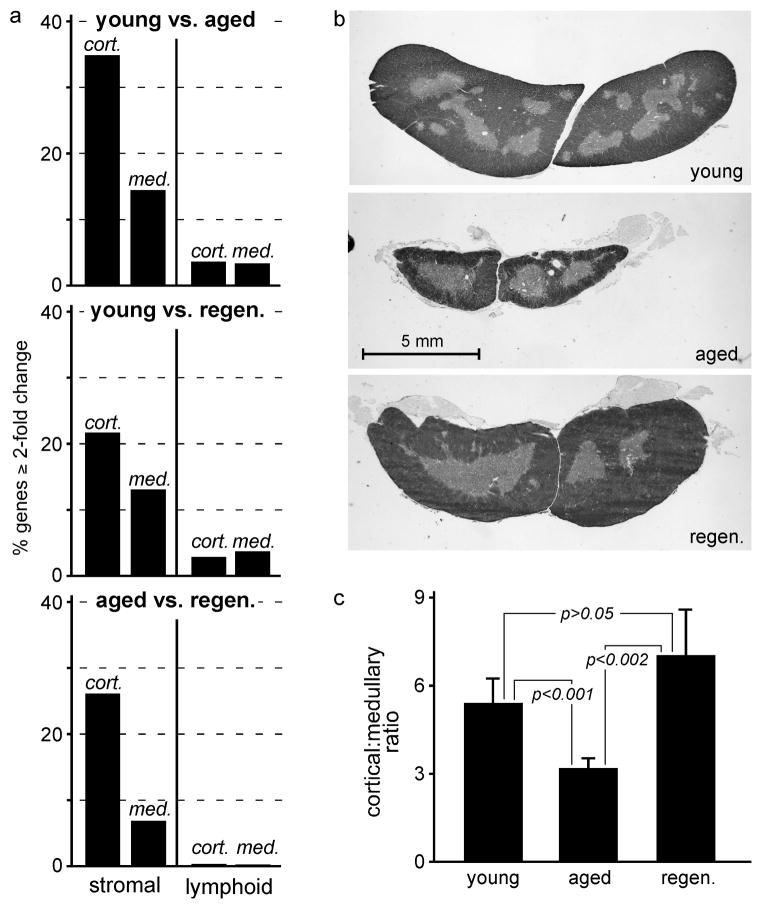
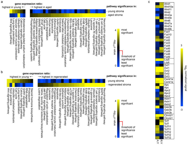
The thymus is the most rapidly aging tissue in the body, with progressive atrophy beginning as early as birth and not later than adolescence. Latent regenerative potential exists in the atrophic thymus, because certain stimuli can induce quantitative regrowth, but qualitative function of T lymphocytes produced by the regenerated organ has not been fully assessed. Using a genome-wide computational approach, we show that accelerated thymic aging is primarily a function of stromal cells, and that while overall cellularity of the thymus can be restored, many other aspects of thymic function cannot. Medullary islet complexity and tissue-restricted antigen expression decrease with age, representing potential mechanisms for age-related increases in autoimmune disease, but neither of these is restored by induced regrowth, suggesting that new T cells produced by the regrown thymus will probably include more autoreactive cells. Global analysis of stromal gene expression profiles implicates widespread changes in Wnt signaling as the most significant hallmark of degeneration, changes that once again persist even at peak regrowth. Consistent with the permanent nature of age-related molecular changes in stromal cells, induced thymic regrowth is not durable, with the regrown organ returning to an atrophic state within 2 weeks of reaching peak size. Our findings indicate that while quantitative regrowth of the thymus is achievable, the changes associated with aging persist, including potential negative implications for autoimmunity.


Figures

Figure 1
Figure 1. Thymic regrowth in response to castration is robust but not durable
Male mice at 11–13 months of age (nominally, 12 months) were castrated, followed by measurement of total cellularity at various intervals, as indicated. Red circles indicate individual thymuses (n = 48 animals total for this figure); the black line indicates a polynomial curve fit to this data. Cellularity exhibited a rapid initial decrease, reaching a nadir at day 3, followed by a period of rapid growth. Growth peaked at approximately 20 days post-castration, at which time cellularity was virtually indistinguishable from that found in 5–6 week-old mice (peak thymus size, data not shown). However, regrowth in response to castration was not durable, and cellularity had degenerated to near the original (pre-castration) value by approximately 2 weeks later.
Figure 2
Figure 2. Aging and regeneration primarily reflect changes in stromal cells, and are dominated by changes in the cortex
Panel a) shows the proportion of all genes (probesets) that were expressed (nominally, signal > median for PLIER signals) in at least one condition (young, aged, or regenerated) in stromal or lymphoid cells from the indicated compartments (cortex, medulla), and that also changed at least 2-fold in the indicated comparisons (young:aged, young:regenerated, aged:regenerated). Changes in cortical cells were dominant, with changes in the cortex dominating those in the medulla. Changes in lymphoid cells were minimal, indicating that the primary changes associated with early atrophy of the thymus are stromal in nature. Panel b) shows typical transverse sections from the mid-point of the thymus of young, aged, and 20 days post-castration mice. Panel c) shows relative quantitation of cortical:medullary area ratios (mean ± s.d.) for 5 thymuses of each type (significance determined by two-tailed Student’s t-test for unpaired samples). Both b) and c) substantiate the conclusion that the most profound changes occurring during both aging and regeneration occur in the cortex. Each experiment was repeated 3–7 times, as described in Materials and Methods, and each experiment contained RNA pooled from multiple mice. Actual numbers of probesets in each bar are as follows: young vs. aged cortical stroma (CS) = 14,914, medullary stroma (MS) = 6663, cortical lymphocytes (CL) = 1895, medullary lymphocytes (ML) = 1728; young vs. regenerated CS = 9940, MS = 6071, CL = 1444, ML = 1853; aged vs regenerated CS = 11,798, MS = 3114, CL = 220, ML = 89.
Figure 3
Figure 3. Medullary complexity and tissue-restricted antigen (TRA) expression decrease with age, and are not restored when the thymus is regrown
Panel a) shows medullary islet counts per lobe for young, aged, and peak regenerated thymuses; bars represent mean ± s.d. for 55 tissue sections from 5 young mice, 109 tissue sections from 6 aged mice, or 81 tissue sections from 4 castrated mice (day 20), while statistical significance was calculated using the two-tailed Student’s t-test for unpaired samples. Medullary islet number decreased approximately 3-fold with age, and did not change even after complete regrowth. Panel b) shows relative expression of TRA in medullary stromal cells from young, aged, and regenerated thymuses; statistically significant changes in expression (two-tailed Student’s t-test for unpaired samples, corrected for multiple testing, see Methods) are indicated by black points, while those that were not statistically significant are indicated in gray. Medullary stroma from aged mice showed substantial changes in TRA expression. The majority of these, representing a broad panel of non-thymic antigens (Suppl. Table 2), were decreased; those that increased exclusively represented B lineage genes, consistent with an increase in the frequency of B cells in the medulla with age (panel c, B220 staining in red, cytokeratin staining in green). The same trend was observed when young medullary stroma was compared to that in the regenerated thymus, with the specific identities of these changed genes almost completely overlapping with that found in young vs. aged. Importantly, there were no significant differences in TRA expression when aged stroma was compared to regenerated, indicating that, like medullary complexity, quantitative regrowth of thymic mass does not translate to a qualitative restoration of TRA expression. Each experiment was repeated 3–7 times, as described in Materials and Methods, and each experiment contained RNA pooled from multiple mice.
Figure 4
Figure 4. Computer learning algorithms distinguish young from aged or young from regenerated stromal gene signatures, but cannot distinguish aged from regenerated
Panel a) shows results of unsupervised two-dimensional clustering of absolute MAS5 values values (log2 signal) for all genes expressed in either young (7 gene chips), aged (3 gene chips), or regenerated (3 gene chips) medulla or cortex, as indicated. All samples from young mice clustered together, and were distinct from aged or regenerated samples. Importantly, aged and regenerated datasets failed to segregate from each other, and were essentially indistinguishable at a global level, despite quantitative changes in organ size. Even when only those genes that underwent a significant (corrected ANOVA p < 0.05) fold change were analyzed (panel b), as opposed to all expressed genes (panel a), the same result was obtained, suggesting that the differences between young and aged thymus persist in the regrown thymus, despite the apparent restoration of cellularity. Each experiment was repeated 3–7 times, as described in Materials and Methods, and each experiment contained RNA pooled from multiple mice.
Figure 5
Figure 5. Biological pathways associated with transcriptional signatures in young, aged, or regenerated cortical stromal cells
Genes represented by the top 20% of cortical stromal signal ratios (young aged, panel a; young regenerated, panel b) were mapped onto KEGG pathways, and pathways were represented at statistically significant levels (Benjamini-Hochberg corrected p value) are shown. Similar to the findings shown in Figs. 3 and 4, pathways representing differences between young and aged were very consistent with those distinguishing differences between young and regenerated, indicating that the regenerated thymus maintains most of the molecular characteristics of the aged thymus, despite an increase in size. This is further illustrated by analysis of individual components of the Wnt signaling pathway, which is the most negatively regulated pathway both during aging (panel a) and in the regrown thymus (panel b). Note that in some cases (in particular, ubiquitin-mediated proteolysis and, to a lesser extent, Wnt signaling), pathways may appear as both higher in young than aged/regenerated, and higher in aged/regenerated than young; this is because different paralogs, and/or positive vs. negative regulators of the pathway, may move in different directions, allowing statistical significance in both cases. A complete description is beyond the limitations of this manuscript, but all relevant data are contained in Suppl. Table 1. Panel c shows relative (normalized to young cortical stroma) gene expression for canonical Wnt pathway components in aged or regenerated stroma; again, the changes seen in regenerated stroma are very similar to those found in aged stroma, indicating the persistence of age related changes after regrowth.

References

    1. Balciunaite G, Keller MP, Balciunaite E, Piali L, Zuklys S, Mathieu YD, Gill J, Boyd R, Sussman DJ, Hollander GA. Wnt glycoproteins regulate the expression of FoxN1, the gene defective in nude mice. Nat Immunol. 2002;3:1102–1108. - PubMed
    1. Besedovsky HO, Sorkin E. Thymus involvement in female sexual maturation. Nature. 1974;249:356–358. - PubMed
    1. Boehm T, Bleul CC. The evolutionary history of lymphoid organs. Nat Immunol. 2007;8:131–135. - PubMed
    1. Brelinska R, Malendowicz LK, Malinska A, Kowalska K. Characteristics of age-related changes in rat thymus: morphometric analysis and epithelial cell network in various thymic compartments. Biogerontology. 2008;9:93–108. - PubMed
    1. Brunelli R, Frasca D, Spano M, Zichella L, Doria G. Gonadectomy in old mice induces thymus regeneration but does not recover mitotic responsiveness. Ann N Y Acad Sci. 1992;673:252–255. - PubMed

Publication types

MeSH terms