. 2012 Feb;80(2):768-77.
doi: 10.1128/IAI.05730-11. Epub 2011 Nov 21.

Toxoplasma gondii triggers release of human and mouse neutrophil extracellular traps

Affiliations

Toxoplasma gondii triggers release of human and mouse neutrophil extracellular traps

Delbert S Abi Abdallah et al. Infect Immun. 2012 Feb.

Abstract

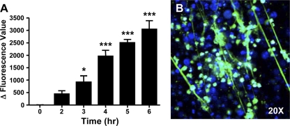
Neutrophils have recently been shown to release DNA-based extracellular traps that contribute to microbicidal killing and have also been implicated in autoimmunity. The role of neutrophil extracellular trap (NET) formation in the host response to nonbacterial pathogens has received much less attention. Here, we show that the protozoan pathogen Toxoplasma gondii elicits the production of NETs from human and mouse neutrophils. Tachyzoites of each of the three major parasite strain types were efficiently entrapped within NETs, resulting in decreased parasite viability. We also show that Toxoplasma activates a MEK-extracellular signal-regulated kinase (ERK) pathway in neutrophils and that the inhibition of this pathway leads to decreased NET formation. To determine if Toxoplasma induced NET formation in vivo, we employed a mouse intranasal infection model. We found that the administration of tachyzoites by this route induced a rapid tissue recruitment of neutrophils with evidence of extracellular DNA release. Taken together, these data indicate a role for NETs in the host innate response to protozoan infection. We propose that NET formation limits infection by direct microbicidal effects on Toxoplasma as well as by interfering with the ability of the parasite to invade target host cells.


Figures

Fig 1
Fig 1
Thioglycolate-elicited mouse neutrophils produce NETs in response to PMA. Purified PMN from LYS-eGFP mice were incubated for 4 h in medium alone (A) or in medium with 600 μM PMA (B). NETs were visualized by staining with DAPI (a) and histone H3 (b). Lysozyme was retained within cells, confirming that NET formation was a regulated event rather than nonspecific cell lysis (c). Merged images are shown in panel d. The scale bar indicates 20 μm. These experiments were repeated at least 3 independent times, with similar results.
Fig 2
Fig 2
Quantitative measurement of PMA and Toxoplasma-induced NET formation. Mouse neutrophils were either stimulated with 600 μM PMA (A) or infected with RH strain tachyzoites at an MOI of 5:1 (B), and NET formation was measured by solubilizing extracellular DNA and measuring Picogreen fluorescence in supernatants as an indicator of double-stranded DNA. Data for both experiments have been corrected to account for the contribution to the fluorescence signal by either cells incubated with medium alone or parasites incubated with medium alone. Experiments were repeated at least 3 independent times, with similar results. ∗, P < 0.05; ∗∗∗, P < 0.005 (relative to time zero).
Fig 3
Fig 3
Release of NETs occurs in a parasite strain-independent manner. Purified mouse neutrophils were added to type I (RH) (A to D), type II (PTG) (E and F), and type III (CTG) (G and H) strains of Toxoplasma, and NET formation was assessed 4 h later by staining for DNA (A, F, and H) and histone H3 (B). Tachyzoites were visualized by staining with Ab to the parasite surface protein SAG-1 (C, E, and G). The insets in panels D, F, and H are merged and expanded images showing the entrapment of parasites within NETs. The scale bars indicate 20 μm. Experiments were repeated at least 3 independent times, with similar results.
Fig 4
Fig 4
NET formation in murine neutrophils does not require invasion. Neutrophils were incubated with Toxoplasma (RH strain at an MOI of 5:1) for 4 h in the presence of cytochalasin D (1 μM). (A) DAPI staining of DNA revealing the presence of NETs. (B) The same image showing parasite SAG-1 staining. (C) Merged image. The merged image in panel C and the expanded area within the red box reveal SAG-1-positive material entrapped within a NET. The scale bar in panel C represents 10 μm. This experiment was performed on three independent occasions.
Fig 5
Fig 5
Parasites are killed in the presence of NETs. (A to D) Neutrophils were incubated with transgenic RH strain parasites expressing Tomato Red fluorescent protein in the presence of cytochalasin D, and 4 h later, cells were stained with the live-cell exclusion DNA dye Sytox Green. (A) DAPI stain; (B) red fluorescence associated with parasites; (C) Sytox Green staining; (D) Merged image. Panel D and the enlarged field show the merged image. Yellow arrows indicate parasites whose nuclei were stained with Sytox Green (nonviable), and red arrows indicate tachyzoites (TZ) that exclude the dye (viable). (E to H) In parallel, the experiment was carried out in the presence of DNase (1 μg/ml). Under this condition, NETs failed to form (E and G), and parasites (F) excluded Sytox Green (G and H). (I) Quantitation of Sytox Green-positive parasites identified by red fluorescence, incubated alone (TZ), in the presence of neutrophils (PMN/TZ), and in the presence of neutrophils with DNase (PMN/TZ/DNase). ∗, P < 0.05. This experiment was performed on three independent occasions.
Fig 6
Fig 6
Decreased parasite viability following incubation with neutrophils. (A) Schematic showing the experimental setup. Parasites were incubated in the presence of cytochalasin D with or without neutrophils for 6 h. The contents of the wells were collected, washed, and added to fibroblast (Fϕ) monolayers to measure parasite infectivity. The formation of plaques, indicative of foci of infection, was visualized by the staining of monolayers with Diff-Quik. (B) Example of fibroblast monolayers in which lysis was allowed to proceed to completion in RH-infected cultures compared in parallel to the same number of tachyzoites coincubated with neutrophils. (C) Plaque formation after the addition of RH and PTG tachyzoites preincubated in the presence or absence of neutrophils. Plaques were enumerated on day 5 (RH) and day 7 (PTG) following inoculation onto fibroblast monolayers.
Fig 7
Fig 7
Differentiated HL-60 cells form extracellular traps in response to Toxoplasma. HL-60 cells were differentiated into neutrophil-like cells with DMSO and then subsequently cultured with T. gondii. (A) Extracellular DNA release relative to medium controls was measured by a Picogreen binding assay. ∗, P < 0.05; ∗∗∗, P < 0.005. (B) Image collected 6 h after culture showing Picogreen-positive NET-like structures induced by Toxoplasma. The experiments were repeated at least 3 independent times, with similar results.
Fig 8
Fig 8
Human peripheral blood neutrophils produce NETs in response to Toxoplasma. (A) Purified neutrophils were incubated with 60 nM PMA or tachyzoites (MOI of 5:1) with or without cytochalasin D (CytD) (1 μM), and the release of DNA was measured over time by using a Picogreen fluorescence assay. Tg, Toxoplasma gondii. ∗, P < 0.05; ∗∗, P <0.01; ∗∗∗, P < 0.005. (B and C) Visual confirmation of NET formation in response to parasites at 4 h postincubation as determined by DNA release (B) in the proximity of parasites (C). (D and E) The merged image (D) and the expanded view (E) show extensive parasite entrapment within NETs. Results are representative of at least 3 independent experiments.
Fig 9
Fig 9
Activation of ERK1/2 in response to Toxoplasma controls NET extrusion. (A) Human peripheral blood neutrophils were incubated in medium alone, with Toxoplasma (Tg) alone, or with the MEK inhibitor U0126. Western blotting was performed to determine the phosphorylation of ERK and p38 MAPK, and blots were stripped and reprobed for total MAPK levels. (B) Images over time from DAPI-stained cells incubated with parasites (top) or with parasites and the ERK inhibitor (bottom). (C) NET formation over time by neutrophils either treated or not treated with the MEK1/2 inhibitor using the Picogreen DNA measuring kit. These experiments were repeated 3 times, with similar results. ∗, P < 0.05; ∗∗, P <0.01; ∗∗∗, P < 0.005; ns, not significant.
Fig 10
Fig 10
(A to F) Neutrophils form NETs in vivo during Toxoplasma infection. Mice were injected intranasally with PBS as a control (A to C) or infected intranasally with PTG strain parasites (D to F). Six hours later, lungs were collected and sectioned, and serial sections were stained with hematoxylin and eosin (HE) (A and D), Ab to myeloperoxidase (MPO) (B and E), and an anti-T. gondii antiserum (TOXO) (C and F). (G) Mice were administered control IgG or neutrophil-depleting MAb 1A8 and then administered PBS or RH strain tachyzoites intranasally. After 6 h, BALF was collected, and dsDNA was measured in cell-free supernatants. (H) BALF cell pellets from the intranasal infection were plated onto fibroblast monolayers, and 5 days later, plaques were enumerated. The experiments were repeated three times, with similar results.

References

    1. Abi Abdallah DS, Egan CE, Butcher BA, Denkers EY. 2011. Mouse neutrophils are professional antigen-presenting cells programmed to instruct Th1 and Th17 T-cell differentiation. Int. Immunol. 23:317–326 - PMC - PubMed
    1. Appelberg R. 2007. Neutrophils and intracellular pathogens: beyond phagocytosis and killing. Trends Microbiol. 15:87–92 - PubMed
    1. Baker VS, et al. 2008. Cytokine-associated neutrophil extracellular traps and antinuclear antibodies in Plasmodium falciparum infected children under six years of age. Malar. J. 7:41. - PMC - PubMed
    1. Ball CJ, King MR. 2011. Role of c-Abl in L-selectin shedding from the neutrophil surface. Blood Cells Mol. Dis. 46:246–251 - PMC - PubMed
    1. Beiter K, et al. 2006. An endonuclease allows Streptococcus pneumoniae to escape from neutrophil extracellular traps. Curr. Biol. 16:401–407 - PubMed

Publication types

Substances

LinkOut - more resources